
Figures & data
Table 1 Baseline Clinical Characteristics of Patients with Renal Involvement
Table 2 Kidney Biopsy Findings of Monoclonal Immunoglobulin-Associated Renal Lesions in 51 NDMM Patients
Figure 1 A patient with light chain cast nephropathy and light chain amyloidosis. (A) showed κ light chain was negative on the arteriolar wall and protein cast (×200); (B) showed λ light chain was positive on the arteriolar wall and strong positive on the protein cast (×200); (C) showed Congo-red positive amyloid on the arteriolar/artery wall and weak positive in the glomeruli (×400); (D) showed PAS-negative protein casts in tubular lumen (arrow, ×200, periodic acid-Schiff staining); (E) showed cell infiltration around the protein casts (arrow, ×200, hematoxylin and eosin staining); (F) showed non-branch fibrils in the subepithelial area of glomeruli (×40,000).
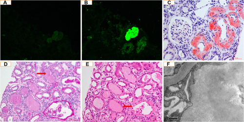
Table 3 Pathological Characteristics of Monoclonal Immunoglobulin-Associated Renal Lesions at Diagnosis
Table 4 Clinical Characteristics of Patients with Kidney Biopsy-Proven Monoclonal Immunoglobulin-Associated Renal Lesions at Diagnosis
Table 5 Treatment and Outcomes
Table 6 Prognostic Factors Associated with Renal Survival
Figure 2 (A) shows renal survival of patients divided according to renal histology. The median renal survival was 6.9 months in the pure MCN group and was not reached in the non-MCN and MCN + other pathologies groups. Log rank p = 0.0093. (pure MCN vs MCN + other pathologies, p = 0.3993; pure MCN vs non-MCN, p = 0.0021; MCN + other pathologies vs non- MCN, p = 0.2163); (B) shows overall survival of patients divided according to renal histology. The median overall survival was 8.7 months in the pure MCN group and was not reached in the non-MCN and MCN + other pathologies groups. Log rank p = 0.0418 (pure MCN vs MCN + other pathologies, p = 0.124; pure MCN vs non-MCN, p = 0.0298; MCN + other pathologies vs non- MCN, p = 0.800).
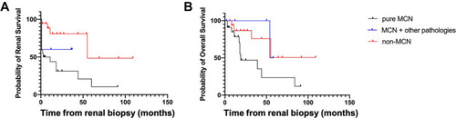
Table 7 Prognostic Factors Associated with Overall Survival