
Figures & data
Figure 1 Cytological specimens of TNs assigned to the AUS/FLUS category of TBSRTC. Hematoxylin and eosin staining, 400× magnification. (A) Cytological atypia. Some cells have mild nuclear enlargement and slight nuclear pleomorphism. (B) Predominant population of microfollicles in an aspirate. (C) Cellular sample composed almost entirely of Hürthle-like cells in a sparse cellular aspirate. Atypia of undetermined significance presenting focal crowded follicular cell clusters with abundant colloid. AUS/FLUS: atypia of undetermined significance/follicular lesion of undetermined significance; TBSRTC: The Bethesda System for Reporting Thyroid Cytopathology.
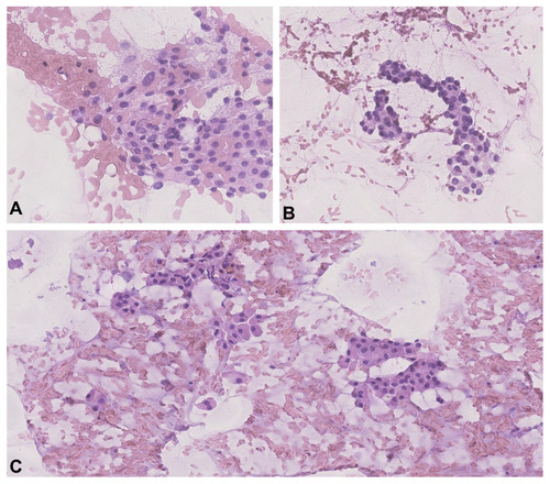
Figure 2 Suspicious ultrasound imaging features of TNs assigned to the AUS/FLUS category in UG-FNAB examination. (A) Transverse US imaging of a TN with microcalcifications (white arrows) without posterior acoustic shadowing. Histopathology revealed PTC. (B) Transverse US imaging of a TN with irregular margins (yellow arrows), microcalcifications (white arrows) and marked hypoechogenicity (blue arrows). Histopathology revealed fvPTC. (C) Transverse US imaging of a TN demonstrating a taller-than-wide shape (brackets). Hypoechogenicity is also presented. Histology revealed PTC. (D) Sagittal US imaging of TN with irregular margins (yellow arrows), microcalcifications (white arrows), cystic components (blue arrows) and hypoechogenicity. Histopathology revealed PTC. AUS/FLUS: atypia of undetermined significance/follicular lesion of undetermined significance; UG-FNAB: ultrasound guided fine needle aspiration biopsy; US: ultrasonography; TN: thyroid nodule; PTC: papillary thyroid cancer; fvPTC: follicular variant of papillary thyroid cancer.
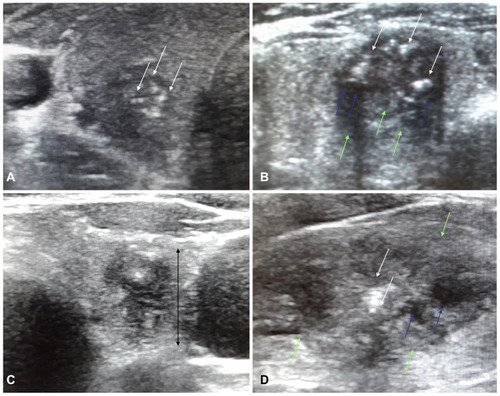
Figure 3 Flowchart of patient selection with individuals finally included in the study. AUS/FLUS: atypia of undetermined significance/follicular lesion of undetermined significance.
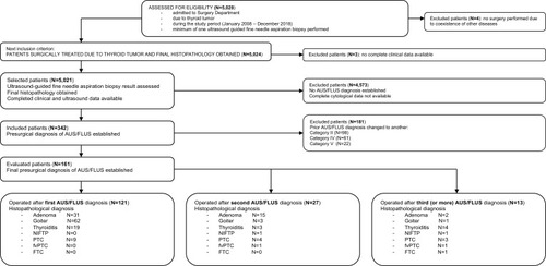
Table 1 Demographic and Clinical Parameters and Ultrasound Features of the 161 Patients with Bethesda Category III (AUS/FLUS)
Table 2 Demographic, Clinical and Ultrasound Characteristics of Three Subgroups of Patients with Bethesda III Category According to Age Parameter. Data are Presented as the Number of Observations (Percent)
Table 3 Logistic Regression Analysis of Ultrasound Variables as Risk Factors of Cancer Presence in Total Group of Patients with Bethesda III Category. Results Were Calculated by Chi-Square Wald Test
Table 4 Logistic Regression Analysis of Age Variable as Risk Factors of Malignancy Presence, High Vascularity and Fast Growth of Thyroid Nodules in Patients with Bethesda III Category. Results Were Calculated by Chi-Square Wald Test