Figures & data
Table 1 Clinical Features of SCLC Patients
Table 2 Detailed Data of Each SCLC Patient
Table 3 Treatment Response
Figure 1 Decrease of tumor marker levels post DEB-BACE treatment. The NSE level (A) and ProGRP level (B) at preoperation, 1st month, 2nd month and 6th month post DEB-BACE treatment in relapsed/refractory SCLC patients.
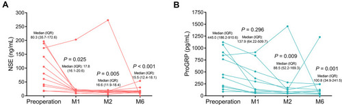
Table 4 QLQ-C30 Score Before and at 2 Months After Treatment
Table 5 Cox’s Regression Analysis of Factors Related to PFS and OS
Figure 2 PFS and OS. The median values of PFS (A) and OS (B) in total patients, and the correlations of PFS (C) as well as OS (D) with age.
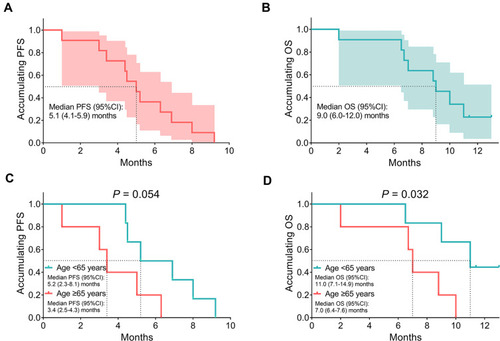
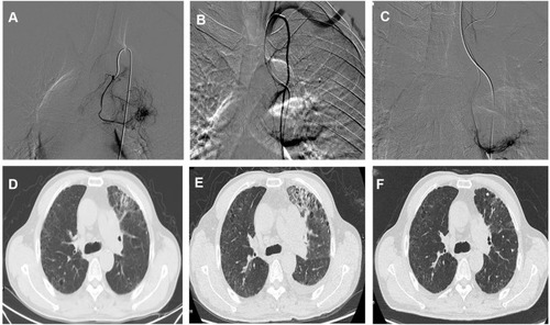
Figure 3 Angiography and chest CT images of a SCLC patient. The images of angiography in the lung during DEB-BACE (A–C), CT image of the reduction of lesion after chemotherapy in the left lung (D), CT image showing a progression of the lesion after chemotherapy (E), and the CT image showing reduction of lesion after DEB-BACE treatment (F).