Figures & data
Figure 1 LncRNA AATBC was significantly up-regulated and its knock-down dramatically inhibited the growth of PCa in vitro and in vivo. (A) Expression of AATBC was detected by qRT-PCR in PCa tissues (n=86) and adjacent normal tissues (n=86). (B) Expression of AATBC was significantly upregulated in PCa cells by qRT-PCR analysis. (C) The validity of AATBC silence was confirmed by qRT-PCR. (D–F) The proliferation and colony formation abilities in PC3 and LNCAP cells of AATBC knockdown were obviously repressive. (G) The apoptosis in AATBC-silenced PC3 and LNCAP cells were disclosed through Annexin V-FITC/PI apoptosis detection. (H–I) Upon downregulation of AATBC expression, the migration and invasion abilities of PC3 and LNCAP cells were measured by transwell assay (scale bars: 100 μm). (J) Knocking down of AATBC significantly shrunk the volume of xenografted tumor in vivo compared to the negative control with transfected with sh-NC. *P<0.05; **P<0.01.
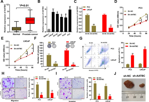
Figure 2 miR-1245b-5p was sponged by AATBC and restrained the progression of PCa. (A and B) Subcellular location of AATBC was determined by nucleus-cytoplasm fractionation and FISH. Scale bars, 20 μm. (C) Starbase database predicted the four miRNAs that had the binding sites to AATBC, and their expression were determined in the PCa cells by qRT-PCR. (D) Pearson correlation analysis showed the passive relationship between miR-1245b-5p and AATBC expression in PCa tissues from our cohort. (E) Predicted binding sites between miR-1245b-5p and AATBC 3ʹ-UTR. (F–H) Biotin-labeled pull-down assay, luciferase assay and RIP assay were used to affirm the interaction between the miR-1245b-5p and AATBC, respectively. (I) The competence of miR-1245b-5p overexpression was validated by qRT-PCR. (J–L) The proliferation and colony formation abilities in miR-1245b-5p overexpressed PC3 and LNCAP cell were significantly augmented by CCK8 and colony formation assay analysis. (M) miR-1245b-5p overexpression significantly promoted the apoptosis of PC3 and LNCAP cells through Annexin V-FITC/PI apoptosis detection. (N and O) miR-1245b-5p overexpression significantly inhibited the migrated and invasive abilities of PC3 and LNCAP cells by transwell assay, respectively. *P<0.05; **P<0.01.
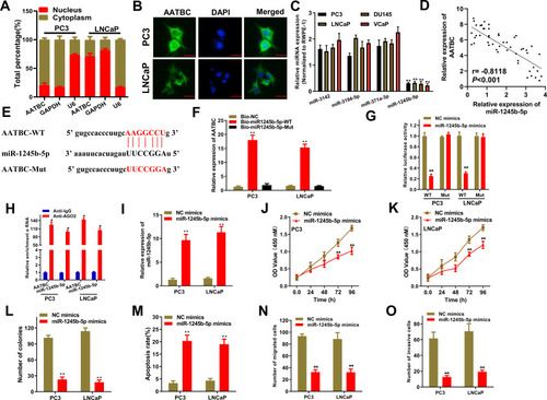
Figure 3 CASK was one downstream target of miR-1245b-5p and boosted the progression of PCa. (A) Venn diagram exhibited the predicted target genes of miR-1245b-5p by the crosstalk of four databases (starbase, miRDB, targetscan and miRWalk), and 5 common genes were selected as the candidate genes. (B and C) qRT-PCR assays confirmed that the expression of CASK could be altered by the overexpression of miR-1245b-5p and silence of AATBC in PC3 and LNCAP cells. (D and E) The mRNA and protein expression of CASK was quantified in PCa cells and normal cells by qRT-PCR and Western blot, respectively. (F) RIP assay justified that AATBC, miR-1245b-5p and CASK could be coexisted in Ago2 pallets. (G) Bioinformatics database predicted the binding site between miR-1245b-5p and CASK. (H and I) Luciferase assay and RNA pull down assay elucidated the interaction between the miR-1245b-5p and CASK above the binding site. (J) The knockdown efficiency of miR-1245b-5p was validated by qRT-PCR. (K and L) CASK mRNA expression and protein levels were determined by RT-qPCR and Western blot in sh-NC, sh-AATBC and sh-AATBC+miR-1245b-5p inhibitors groups. (M) The knockdown competence of CASK was validated by qRT-PCR. (N–Q) The influence of biological function including colony formation (N), apoptosis (O), migration (P) and invasion (Q) were determined in PCa cells with the silence of CASKC. **P<0.01.
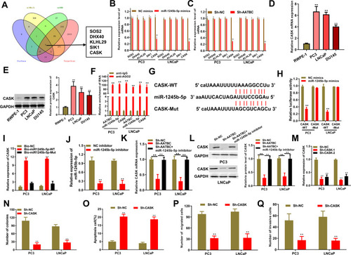
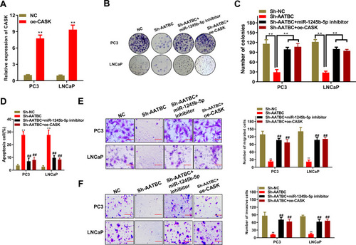
Figure 4 AATBC advanced PCa cell progression by the regulation of miR-1245b-5p/CASK axis. (A) The efficiency of CASK overexpression was manifested by RT-qPCR. (B and C) Colony formation was carried out to weigh cell proliferation competence in PCa cells of AATBC depletion plus miR-1245b-5p downregulation or CASK overexpression. (D) The apoptosis in PCa cells of AATBC depletion plus miR-1245b-5p downregulation or CASK overexpression was determined by the analysis of Annexin V-FITC/PI apoptosis detection kit. (E and F) The migratory and invasive ability of PCa cells of AATBC depletion plus miR-1245b-5p downregulation or CASK overexpression were appraised by transwell assay (scale bars: 100 μm). **P<0.01, vs sh-NC. ##P<0.01, vs sh-AATBC.