Figures & data
Figure 1 The expression level of zinc finger protein 704 (ZNF704) in fresh chondrosarcoma tissues and the paired adjacent non-tumor tissues. (A) Comparison of differences in the ZNF704 mRNA levels between 6 cases of chondrosarcoma tissues and the paired adjacent non-tumor tissues. CS, chondrosarcoma tissues; N, the paired adjacent non-tumor tissues. **P<0.01 by Student’s t test. (B) Representative immunoblotting showed the protein expression of ZNF704 in CS tissues and the paired adjacent non-tumor tissues (Left Panel). Quantitative data revealed that the ZNF704 protein expression in CS tissues was significantly higher than that in the paired adjacent non-tumor tissues (Right Panel). *P<0.05 by Student’s t test.
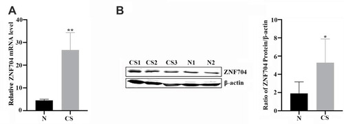
Table 1 The Protein Expression Level of ZNF704 in Human Cartilage Tumors
Figure 2 Prognostic value of ZNF704 in chondrosarcoma cases. (A and B) Representative immunostaining of ZNF704 were shown in human cartilage tumors. Osteochondroma with low nucleus immunostaining of ZNF704 (A). High (B) nucleus immunostaining of ZNF704 were detected in chondrosarcoma tissues. (C) Prognostic significance of ZNF704 in 63 patients with chondrosarcoma. Kaplan-Meier overall survival curves and the Log rank test of 63 cases of chondrosarcoma patients based upon the status of ZNF704 expression were conducted, and unravelled that high ZNF704 expression remarkably correlated with poor prognosis (P=0.01).
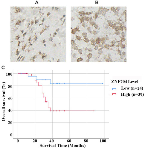
Table 2 Associations Between ZNF704 Expression and the Clinicopathological Characteristics in 63 Patients with Chondrosarcoma
Table 3 Multivariate Analysis of Various Prognostic Parameters in 63 Patients with Chondrosarcoma by the Cox Regression Model
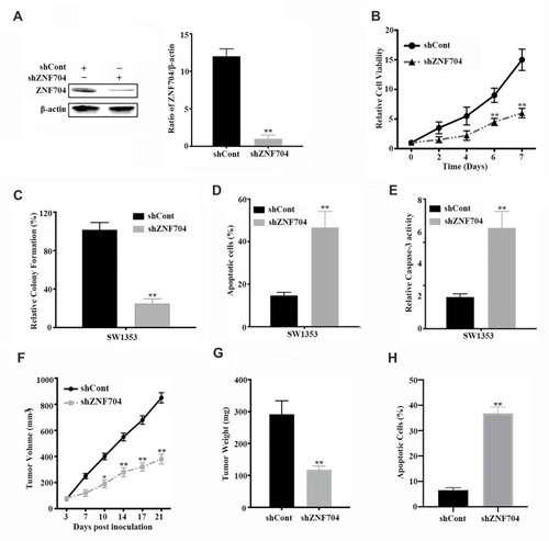
Figure 3 Knockdown of ZNF704 inhibits chondrosarcoma cell proliferation and induces apoptosis in vitro and in vivo. (A) ZNF704 was markedly reduced by shRNA against ZNF704. SW1353 cells transfected with control shRNA (shCont) or shRNA against ZNF704 (shZNF704) were subjected to immunoblotting (Left Panel). The histograms shown the gray intensity analysis to the bands of immunoblotting (Right Panel, **P <0.01). (B and C) The inhibitory effects of shRNA against ZNF704 on cell viability and colony formation. Stable SW1353 cell clones with shCont or shZNF704 were incubated for the indicated time, and then cell viability (B) and colony formation (C) were detected by CCK-8 and clonogenic assay respectively, as indicated in Material and Methods. Data are plotted as the mean ± SD (*P <0.05, **P <0.01). (D) Quantification of the apoptotic cell population by flow cytometry. Stable SW1353 cell clones with shCont or shZNF704 were incubated for 48 hours. Apoptotic cells were measured by FITC-Annexin V and PI staining followed by flow cytometry analysis. Knockdown of ZNF704 induced a larger subset of apoptotic cells compared with shCont. n = 3 repeats with similar results; **P<0.01 by Student’s t test. (E) Quantification of the pro-apoptotic caspase 3 activity. Stable SW1353 cell clones with shCont or shZNF704 were incubated for 48 hours. Caspase-3 activity was measured and revealed that the pro-apoptotic caspase 3 activity was significantly increased after knockdown of ZNF704. n=3 repeats with similar results. **P <0.01 by Student’s t test. (F and G) Knockdown of ZNF704 suppressed tumor growth in a nude mouse xenograft model. Stable SW1353 cell clone with shCont or shZNF704 were implanted into nude mice via subcutaneous injection. Tumor volume (F) and tumor weight (G) of stable SW1353 cell clones for 21 days, respectively, indicated that ZNF704 knockdown slowed down tumor growth in nude mice. n=5, Data are plotted as the mean ± standard deviation (*P<0.05, **P<0.01). (H) Quantification of apoptotic cells using TUNEL assay in SW1353 chondrosarcoma xenografts. The average percentage of TUNEL-positive cells in tumors isolated from shZNF704 were significantly higher than those in the shCont group. n=5, **P<0.01.