Figures & data
Figure 1 Schematic representation of the anatomy of lateral chest wall perforator flaps (CWPF) and the blood supply in relation to breast. T: tumour with outer circle representing the wide local excision; 1) lateral CWPF; 2) latissimus dorsi muscle; 3) lateral thoracic vessels; 4) lateral Intercostal vessels.
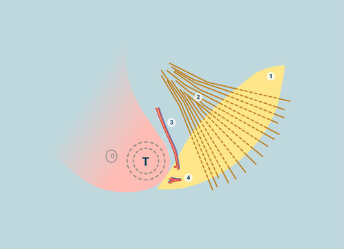
Table 1 Distribution of the Clinic-Pathological and Treatment Parameters
Table 2 Details of the Median Tumor Size in Relation to the Bra Cup
Figure 2 One-stage LICAP flap reconstruction (A) 43-year old with 40 mm cancer in the lower outer quadrant of the right breast. Pre-op marking for tumor location and lateral CWPF. The solid vertical line (white arrow) denotes surface marking for lateral thoracic artery (LTA) and stars represent lateral intercostal artery perforators (LICAP). The dotted lines are along the lateral border of pectoralis major and anterior border of latissimus dorsi muscle. (B) Intra-operative picture showing the flap dissected (arrow points towards head with patient in lateral position). (C) 4 weeks post-op with scar on the lateral chest wall. Patient had chemotherapy after surgery. (D) Appearance and symmetry of breasts 4 years after radiotherapy. (E) Appearance of scar 4 years later.
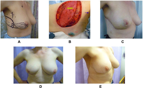
Table 3 Tumor Characteristic of Patients Undergoing One-Stage and Two-Stage Approaches
Figure 3 Flowchart depicting axillary lymph node status along with axillary treatment.
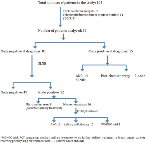
Figure 4 Cancer outcomes in the study (Consort format).
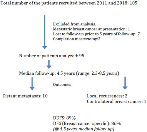
Figure 5 Patient before (A) and after (B) contralateral breast augmentation to address asymmetry after PBR with CWPF. The augmentation was performed 2 years after completion of cancer treatment.
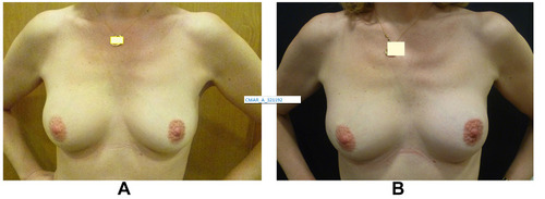
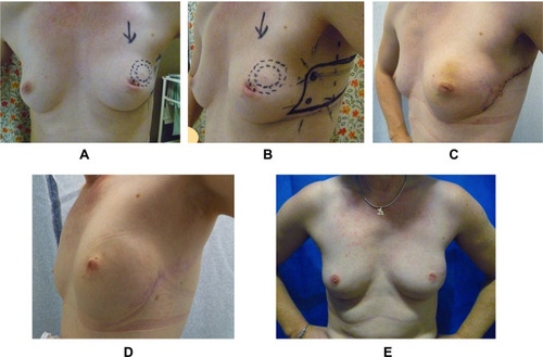
Figure 6 Pre-op and post-op photographs of PBR with lateral CWPF in a slim patient with very small breasts. 42-year old with 20 mm cancer in the upper outer quadrant of left breast with an “A” cup. (A) Pre-operative photograph. (B) Pre-op marking for CWPF. (C) 2 weeks after surgery. (D) 1 year after treatment. (E) 3 years after radiotherapy on right side. Patient had chemotherapy after surgery.