Figures & data
Figure 1 The pathological manifestations of pseudomyxoma peritonei. (A) low-grade mucinous carcinoma peritonei (HE, ×200); (B) high-grade mucinous carcinoma peritonei (HE, ×200); (C) high-grade mucinous carcinoma peritonei with signet ring cells (HE, ×400); (D) MUC2 positive (IHC, ×400; MUC2, clone MRQ-18, catalog number ZM-0392); (E) MUC5AC positive (IHC, ×400; MUC5AC, clone MRQ-19, catalog number ZM-0395); (F) PAS staining of mucus, pink area in the left image (HE, ×200). The staining turned negative when incubated with salivary amylase. Unpublished data from the authors’ group.
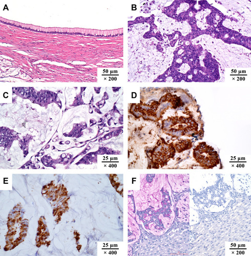
Figure 2 Schematic diagram of MUC2 gene. MUC2 gene is located in chromosome 11p15.5.
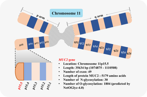
Figure 3 Schematic diagram of the domains in MUC2 amino acid chain.
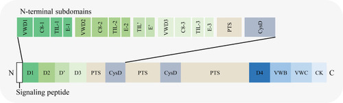
Figure 4 Schematic diagram of MUC2 transcription, translation, and post-translational modification. (A) MUC2 mRNA synthesis in the nucleus; (B) In the ribosome, mRNA is translated to form a polypeptide chain with a length of 5179 amino acid residues; (C) In the endoplasmic reticulum, the N-terminus and C-terminus of the peptide chain were N-glycosylated and dimerized through a disulfide bond; (D) In the Golgi apparatus, the dimerized MUC2 are further O-glycosylated and multimerized via disulfide bond.
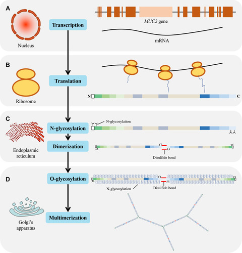
Figure 5 Schematic diagram of N-glycosylation and O-glycosylation sites in MUC2. (A) N-glycosylation; (B) O-glycosylation.
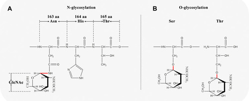
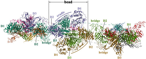
Figure 6 The schematic diagram of the quaternary structure of the MUC2 head. Reproduced from the RCSB PDB database (https://www.rcsb.org/), PDB ID: 7A5O. Sehnal D, Rose AS, Koča J, Burley SK, Velankar S. Mol*: towards a common library and tools for web molecular graphics. Proceedings of the workshop on molecular graphics and visual analysis of molecular data. Brno, Czech Republic: Eurographics Association; 2018:29–33.Citation30