Figures & data
Figure 1 The chest CT outcomes during the whole treatment. (A) (A1–A3) Chest CT reveals primary lung cancer and metastases (red arrow); (B) tumor was PR after 2 cycles chemotherapy and gefitinib; (C) tumor was SD after 2 cycles chemotherapy and gefitinib again; (D) tumor was PD for taking gefitinib only; (E) tumor was PR again after 4 cycles the same chemotherapy and osimertinib; (F) tumor was PD again for taking osimertinib only. (G) Tumor was PR again after 2 cycles anlotinib.
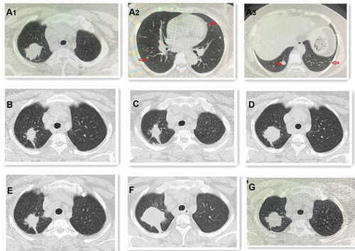
Figure 2 (A) H-E staining shows that SCC (blue arrow) presents a solid sheet-like arrangement. The cell cytoplasm is red stained, the nucleus is large, nucleoli and mitosis, no intracellular keratinizing and keratinizing beads are seen, but AC (red arrow) is adenoid, cord-like or solid arrangement. The cell cytoplasm is reddish or translucent, the nucleus is large and irregular, and nucleoli and mitosis can be seen. (x200). SCC accounts for about 10% and AC accounts for about 90% of the tumor. IHC staining shows: (B) CK5/6 and (C) P40 are positive in SCC components. (x200) (D) TTF-1 and (E) Napsin-A are positive in AC components. (x200) (F) Ki-67 index is about 70% in SCC (blue arrow) and about 30% in AC (red arrow). (x200).
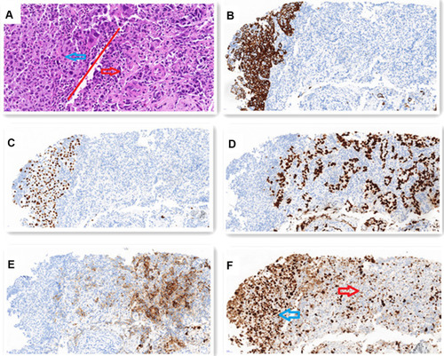
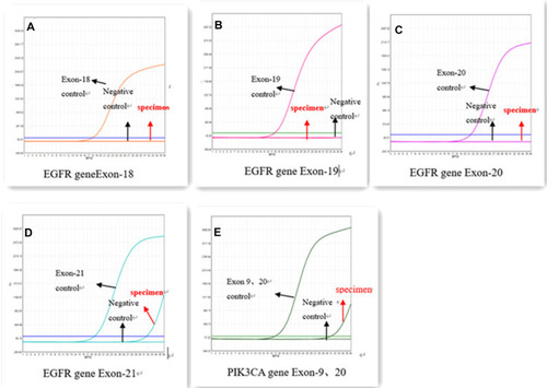
Figure 3 ARMS-PCR gene sequencing illustrated EGFR 18 (A), EGFR 19 (B), EGFR 20 (C) are negative, EGFR 21 (L858R) (D) and PIK3CA (H1047R/E545K) (E) concurrent mutations in AC component.