
Figures & data
Figure 1 Patient flow diagram.
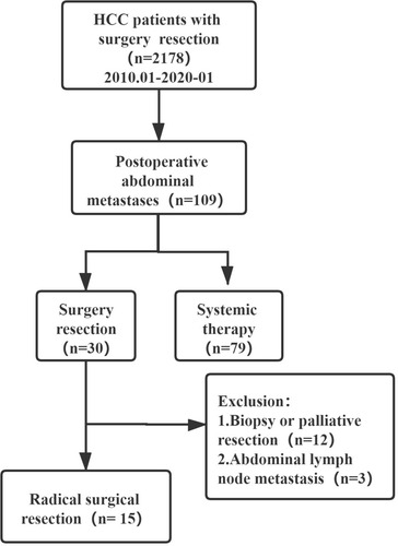
Table 1 Clinicopathologic Characteristics of the 15 Patients with Resected Peritoneal Dissemination
Table 2 Laboratory Data of the 15 Patients with Resected Peritoneal Dissemination
Table 3 Pathological Characteristics of the First Surgery
Figure 2 Clinical picture of patient 1. (A) Giant metastatic tumor of mesentery; (B) Computed tomography (CT) images; (C) Tumor section specimen; (D) Arterial dominant phase of CT shows arterial enhancement of nodule measuring 10.0 cm in diameter.
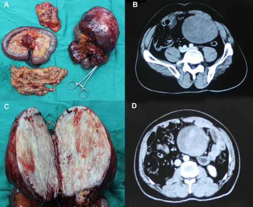
Figure 3 Clinical picture of patient 4. (A) Giant metastatic tumor of omentum; (B) Horizontal plane of magnetic resonance images; (C) Tumor section specimen; (D) Coronal plane of magnetic resonance imaging.
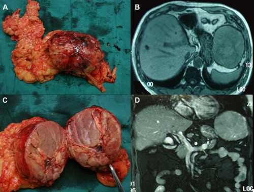
Table 4 Postoperative Survival of the Patients
Figure 4 The survival rate after resection of the abdominal metastases.
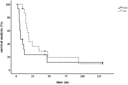
Table 5 Median Survival of Abdominal Metastases of Hepatocellular Carcinoma in the Literature