Figures & data
Figure 1 Regulation of autophagy signaling pathways. Autophagy is a complex degradation process involving the following key steps: (A) initiation; (B) nucleation; (C) maturation; (D) fusion; (E) degradation. During initiation, low ATP, hypoxia, and amino acid deficiency lead to AMPK activation or mTOR inhibition, and the ULK complex forms. The ER membrane breaks off to form phagophores. In the starvation state, JNK1-mediated phosphorylation of BCL2 is blocked. BECN1 separates from BCL2 to form the Class III PI3K complex. BECN1 in the Class III PI3K complex interacts with the ER and participates in double-membrane nucleation to form a phagophore, which contains abnormal proteins and damaged organelles. ATG5 is conjugated with ATG12 and forms a complex with ATG16L, which is involved in phagophore elongation. After LC3 processing, it is inserted into the extended phagophore to form a mature autophagosome. Then, the autophagosome fuses with a lysosome to form the autolysosome and degrade the contents.
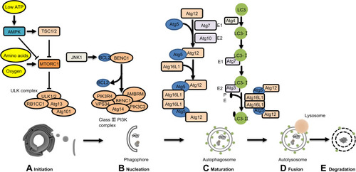
Figure 2 Role of autophagy regulatory factors in pancreatic tumorigenesis in mice with mutant KRAS.
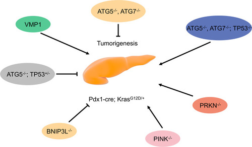
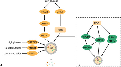
Figure 3 Autophagy maintains the metabolism and function of PDAC cells. Autophagy pathways are modulated by different metabolic conditions (eg, oxidative stress, low glucose, and low amino acids) in which cellular components are degraded. In this process, bioenergy intermediates are reused, thereby promoting cell survival.