
Figures & data
Figure 1 Imaging features of the patient’s lymphoma. (A) Ultrasonography demonstrates a focal area of hypoechogenicity with hypervascularity in the left testis. (B and C) Post-orchiectomy PET/CT shows an FDG-avid lesion (green arrow) in the right testis. (D) PET/CT-based CR was achieved after first-line immunochemotherapy. (E and F) At the first follow-up examination, MRI horizontal and frontal views show a new lesion (red arrow) involving right basal ganglia and pons. (G and H) MRI horizontal and frontal views show that the new lesion disappeared after the first cycle of chemo-free treatment.
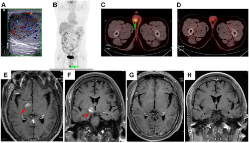
Figure 2 Immunohistochemical features of the patient’s tumor. The lymphoma cells were positive for CD20, CD79a, Mum-1 (dim), BCL-6, BCL-2, and C-MYC, and negative for CD10. Ki-67 proliferation index was about 90%. Original manifestation 400×.
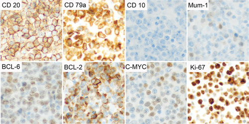
Figure 3 Treatment timelines of the patient.
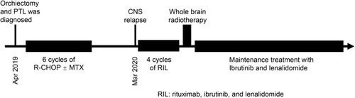
Table 1 Reported PTL Studies
Table 2 Important Molecular Aberrations in PTL
Table 3 Prognostic Factors for PTL