84
Views
0
CrossRef citations to date
0
Altmetric
Review

When to perform positron emission tomography/computed tomography or radionuclide bone scan in patients with recently diagnosed prostate cancer

, , &
Pages 123-131 | Published online: 25 Jun 2013

Figures & data

Figure 1 (AE) A 76-year-old patient with recently diagnosed prostate cancer (prostate-specific antigen level, 18 ng/mL) underwent bone scan and positron emission tomography (PET)/computed tomography (CT) using radiolabeled choline for staging purposes. Bone scan in anterior (A) and posterior view (B) shows an area of mild radiopharmaceutical uptake corresponding to the left ischiatic bone (arrow). This finding was more evident by using single-photon emission computed tomography imaging (C).

Notes: Axial CT (D) and radiolabeled choline PET/CT (E) images showed the presence of increased radiopharmaceutical uptake corresponding to an osteosclerotic lesion located in the left ischiatic bone (arrows). This finding was highly suspicious for skeletal metastasis. Biopsy confirmed the presence of a bone metastasis by prostate cancer.

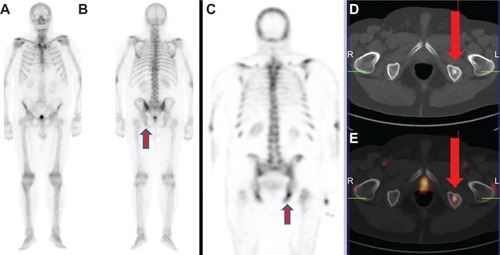
Figure 1 (A–E) A 76-year-old patient with recently diagnosed prostate cancer (prostate-specific antigen level, 18 ng/mL) underwent bone scan and positron emission tomography (PET)/computed tomography (CT) using radiolabeled choline for staging purposes. Bone scan in anterior (A) and posterior view (B) shows an area of mild radiopharmaceutical uptake corresponding to the left ischiatic bone (arrow). This finding was more evident by using single-photon emission computed tomography imaging (C).Notes: Axial CT (D) and radiolabeled choline PET/CT (E) images showed the presence of increased radiopharmaceutical uptake corresponding to an osteosclerotic lesion located in the left ischiatic bone (arrows). This finding was highly suspicious for skeletal metastasis. Biopsy confirmed the presence of a bone metastasis by prostate cancer.