Figures & data
Table 1 Literature Review of Studies Reporting Cervical LNM from CNS Tumors
Figure 1 Flow chart showing the study selection process for our systematic review.
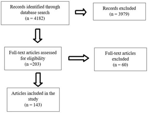
Figure 2 Male, 60 years old, cervical lymph node metastases (arrows) from a GBM. MRI axial T2-weighted sequences (A and E), MRI axial contrast-enhanced T1-weighted sequences (B and F), CT scans axial plane (C and G) and Fused Positron Emission Tomography- Computed Tomography axial plane (D and H). Observe the lymph nodes (arrows) in the right IIB (A–C) and V right level (E–G). Also, notice the increased uptake of fluorodeoxyglucose (FDG) in those lymph nodes (arrows in (D) and (H)).

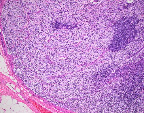
Figure 3 Microphotograph demonstrating an anaplastic ependymoma within a cervical lymph node. (Hematoxylin and Eosin, X200).