Figures & data
Figure 1 Rash: on admission.
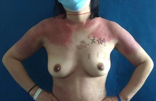
Figure 2 Breast ultrasound showed a 1.5×2.0 × 1.4cm mass in the left breast.
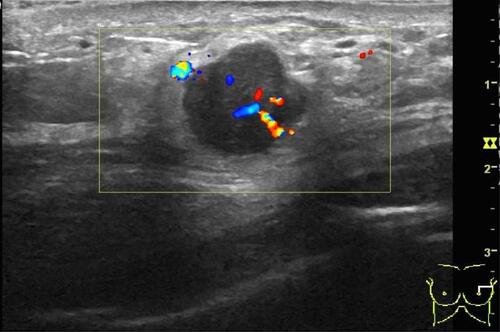
Figure 3 Breast MRI.
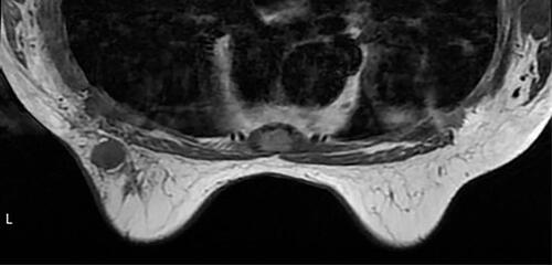
Figure 4 Histological examination shows invasive ductal carcinoma (H-E, original magnification, × 100).
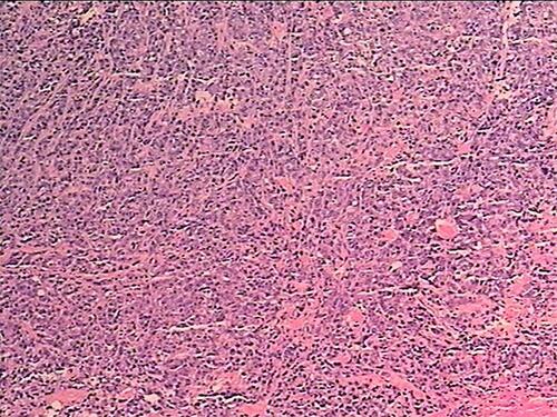
Figure 5 1 week after breast cancer surgery.

Figure 6 Postoperative PET/CT of the breast.
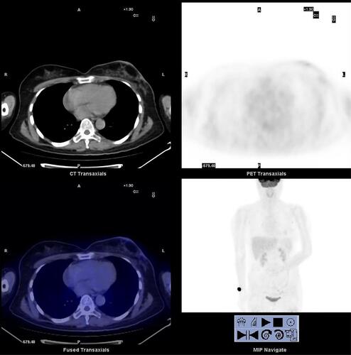
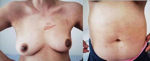
Figure 7 1 year after breast cancer surgery.