
Figures & data
Figure 1 CC pathology and 18F-FDG PET/CT images. (A and B) Clinical responses of the patient to DC+ICI treatment: PR. (C) HE-stained section of the tumor showed extensive hemorrhage and necrosis at low magnification, the biphasic feature of mixed cytotrophoblasts (black triangle) and syncytiotrophoblasts (black arrow) and nuclear division in tumor cells (star) at high magnification. In some regions, tumors are mainly composed of mononuclear cytotrophoblasts, and syncytiotrophoblasts are few and inconspicuous, which is easy to be misdiagnosed as poor-differentiated carcinoma. Immunohistochemical markers provided further support for the diagnosis of CC.
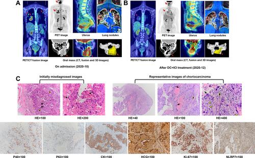
Figure 2 Tumor microenvironment of this patient. (A) Targeted next-generation sequencing analysis (483-gene panel) showed high tumor mutation burden (33.571 muts/MB). (B) Multiplex immunofluorescence staining results.
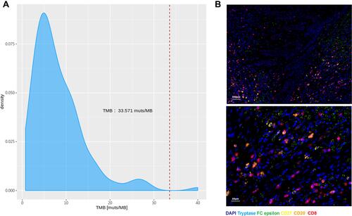
Figure 3 DNA polymorphic analysis. (A–D) Genetic profiles of 21 highly polymorphic short tandem repeats (STRs) from tumor, the patient and spouse were compared. At 7/21 valid loci (D19S433, D7S820, D8S1179, TPOX, Penta E, D12S391, FGA) examined, tumor sample was found to contained both the maternal and paternal allele, demonstrating its gestational origin. Y-axis: fluorescence intensity of the labeled product. X-axis: allelic sizes in base pairs, such that the number stands for repeat size in each short tandem repeat locus and can be highly variable among individuals.
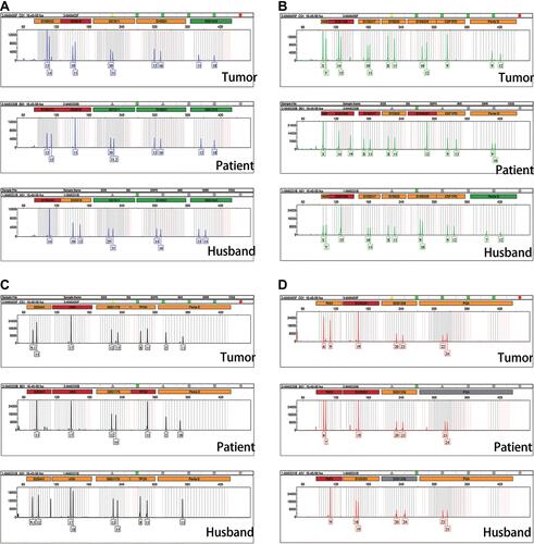
Figure 4 Clinical course. The patient underwent gingival resection, relapse, and was transferred to our hospital to receive 2 cycles of DC+ICI treatment, followed by 3 cycles of FEVA chemotherapy. Pathologic images revealed the biphasic growth pattern of cytotrophoblasts (yellow triangle) and syncytiotrophoblasts (yellow arrow). Dynamic observation of lesions imaging (red arrow) and β-HCG levels were shown below.
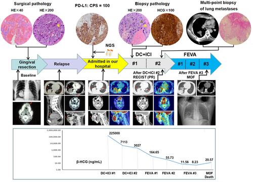
Table 1 Cases of Oral Metastasis by Choriocarcinoma with Survival Data in Reviewed Literatures