Figures & data
Figure 1 Four types of bronchial cutoff signs. (A) Type I: the lesion grows along the outline of the bronchus with a noted truncation of the proximal lumen (red arrow). (B) Type II: bronchus (red arrow) is obstructed abruptly by the lesion. (C) Type III: bronchus (red arrow) penetrates into the lesion with tapered narrowing and interruption. (D) Type IV: bronchus (red arrow) runs around the periphery of the lesion and is displaced, compressed, and narrowed by it.
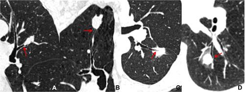
Table 1 Clinical Characteristics of the Studied Patients
Table 2 CT Features of Different Nodules
Table 3 Binary Logistic Regression Analysis of the Clinical and CT Characteristics of Studied Patients
Figure 2 Differentiation between peripheral small-cell lung cancer (pSCLC) and peripheral non-small cell lung cancer (pNSCLC). Axial computed tomography (CT) image in a 76 year old male with pSCLC shows an irregular nodule with homogeneous density and smooth margin (A), 99 days later, the nodule size increases obviously and the right hilar lymphadenectasis is detected simultaneously (red stars) on contrast enhanced CT image (B). Axial and reconstructed CT images in a 73 year old female with pNSCLC show an oval nodule with heterogeneous density, spiculation (red arrows) (C), and pleural indentation (red arrow) (D).
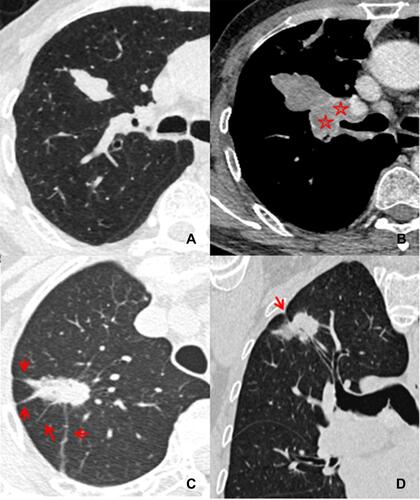
Figure 3 Differentiation between pSCLC and benign lung tumors (pBLT). Axial and reconstructed CT images in a 69 year old female with pSCLC show an oval nodule with a smooth margin, homogeneous density (A) and bronchial cutoff sign (red arrow) (B). Axial CT images in a 51 year old female with pBLT show a round nodule with smooth margin (C), and homogeneous density (D).
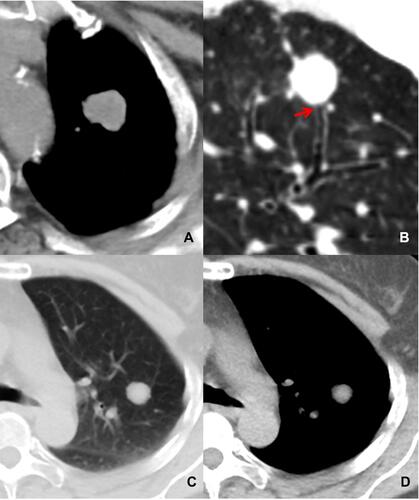
Figure 4 Differentiation between pSCLC and inflammatory lesions (pIL). Axial and reconstructed CT images in a 78 year old male with pSCLC show a round nodule with smooth margin, homogeneous density (A), and the bronchial cutoff sign (red arrow) is also detected (B). Axial and reconstructed CT images in a 46 year old female with pIL show a round nodule with spiculation (C) and peripheral GGO (D) but without bronchial cutoff sign (D).
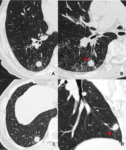
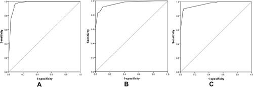
Figure 5 Receiver operating characteristic curves of the pSCLC predictive model established by the independent clinical and CT characteristics mentioned above. (A) performance of the pSCLC predictive model from pNSCLC; (B) performance of the pSCLC predictive model from pBLT; (C) performance of the pSCLC predictive model from pIL.