
Figures & data
Table 1 Summary of Pivotal Trials with Anti-PD1 Antibody Plus Chemotherapy for HER2-Negative AGC
Table 2 Regulatory Approval and Recommendation of Each Region About First-Line Chemotherapy Plus Nivolumab.Citation2,Citation28,Citation29
Table 3 Viewpoints of Each Stance About the Adaptation of Chemotherapy Plus Nivolumab as a First-Line Treatment
Figure 1 Patients’ flow after the first visit from November 2021 to June 2022 in National Cancer Center Hospital East.
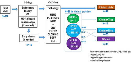
Figure 2 CT scan in case 1. (A) Before the start of nivolumab plus SOX. (B) After the third course of nivolumab plus SOX. Explanations: Arrows in upper pictures show liver metastasis; Arrows in lower pictures show bilateral ovarian metastasis.
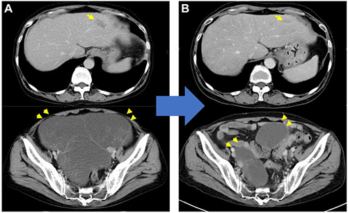
Figure 3 Upper gastrointestinal endoscopy and CT in case 2. (A) Upper gastrointestinal endoscopy before chemotherapy. (B and C) CT revealed interstitial pneumonia and lymph node and lung metastases before chemotherapy. (D–F) CT showed mediastinal and subcutaneous emphysema after two courses of pembrolizumab, while interstitial pneumonia was almost stable and the metastatic lesions had maintained shrinkage. Explanations: Yellow arrows in (C and F) show lymph node metastasis; White arrows in (C and F) shows liver metastasis.
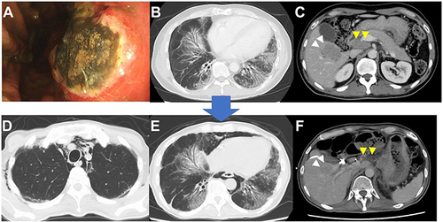
Table 4 Overview of Ongoing Pivotal Clinical Trials, Including ICI as a First-Line Treatment in HER2-Negative AGC