
Figures & data

Figure 1 Representative of patient’s PET imaging. (A and B) PET scan images revealed that FDG uptake was high in multiple lymph nodes (SUV max: 9.4) and bones (SUV max: 6.2), posterior wall of nasopharynx (SUV max: 6.5), bilateral ethmoid sinuses (SUV max: 5.0), bilateral testicle and right spermatic cord (SUV max: 7.3) and subcutaneous nodules on bilateral cheeks (SUV max: 2.8) before therapy. (C and D) PET/CT evaluation was carried out before Protocol M of CCCG-LBL-2016 beginning, and it showed complete remission. (E and F) Repeat PET/CT scan more than five months after transplantation showed normal metabolic activity. FDG, fluorodeoxyglucose; PET, positron emission tomography.
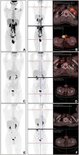
Figure 2 Morphologic features of the skin lesion. (A) The core needle biopsy specimen is extensively replaced with tumor cells characterized by nucleoli and scant cytoplasm (H&E, 400x). (B–D) Immunohistochemical stains show that tumor cells were positive for CD20 ((B), 100x), PAX5 ((C), 100x), and TdT ((D), 100x). (E) Immunohistochemical stains show that tumor cells were negative for CD34 (100x). (F) About 90% of the neoplastic cells display nuclear Ki67 staining (100x). TDT, terminal deoxynucleotidyl transferase.
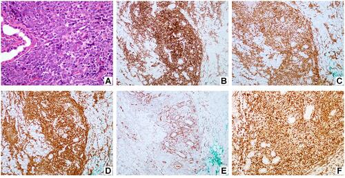
Figure 3 FISH analysis was approached on suspension samples in interphase cells of fresh swelling lymph node specimen using commercial probe (LSI BCR/ABL DS probe, Vysis). (A) Cells with a signal pattern of 1 red, 1 green, and 2 yellow (fusion signal) are positive for BCR/ABL1 rearrangement. (B) Cells with 2 red and 2 green are normal. FISH, Fluorescence in situ hybridization.
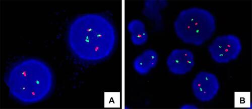
Table 1 Summary of Previous Case Reports