Figures & data
Figure 1 (A and B) Baseline CT images. The red arrow in figure A indicates tumors and the red arrow in figure B indicates lymph nodes. (C and D) Lung mass and nodal metastasis achieved PR after four cycles of AP (albumin paclitaxel 400 mg and carboplatin AUC 5) and sugemalimab 1200 mg. The red arrow in figure C indicates tumors and the red arrow in figure D indicates lymph nodes. (E and F) Lung mass achieved SD, and nodal metastasis achieved CR after five cycles of 1200 mg of sugemalimab. The red arrow in figure E indicates tumors and the red arrow in figure F indicates lymph nodes.
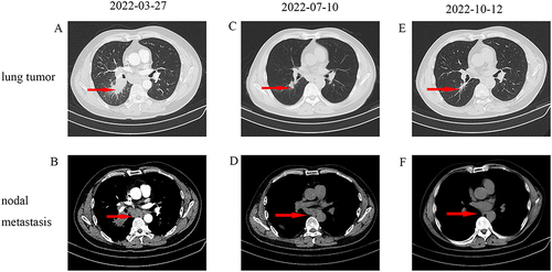
Figure 2 (A) Microscopic appearances of the pulmonary tumor showed lung SCC (HE staining x100). (B–D) Immunohistochemical staining for P40(+), Syn (-) and CD56(-), which are the optimal immunohistochemical markers for SCC (HE staining x100). (E) Microscopic appearances showed transformed SCLC (HE staining x100). (F–H) Immunohistochemical staining for P40(-), Syn (+) and CD56(+), which are the optimal immunohistochemical markers for transformed SCLC (HE staining x100).
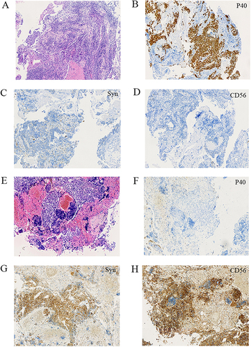
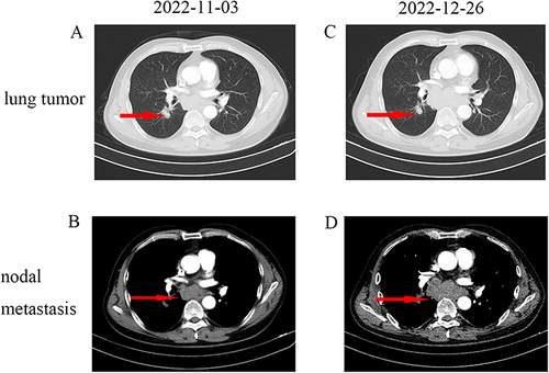
Figure 3 (A and B) Lung mass and nodal metastasis were evaluated as PD compared with CT (12 October 2022). The red arrow in figure A indicates tumors and the red arrow in figure B indicates lymph nodes. (C and D) The lung mass achieved SD, and nodal metastasis achieved PD after two cycles of etoposide combined with cisplatin. The red arrow in figure C indicates tumors and the red arrow in figure D indicates lymph nodes.