
Figures & data
Figure 1 Prediction model of hMCT1 (a) and hMCT4 (b).
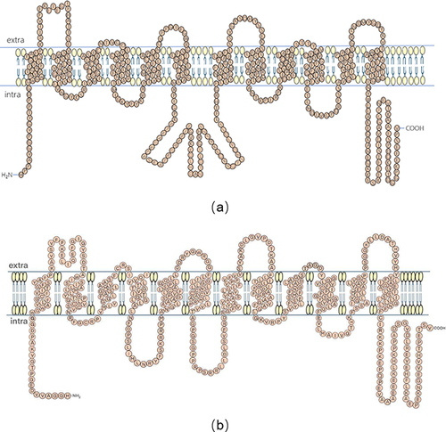
Figure 2 Glucose metabolic process. Glucose enters cells via glucose transporters (GULT). Many enzymes are involved in the process of anaerobic glycolysis. The production of pyruvate from glucose is catalyzed by three key enzymes: hexokinase (HK), 6-phosphofructokinase 1 (PKF1) and pyruvate kinase (PKM). Pyruvate is catalyzed by lactate dehydrogenase A (LDHA) to lactate. The tricarboxylic acid cycle (TCA) process is catalyzed by several key enzymes: citrate synthase (CS), isocitrate dehydrogenase (IDH) and ketoglutarate dehydrogenase complex (KGDHC). Blue areas (eg, red skeletal muscle, heart and neurons) and green areas (eg, white skeletal muscle fibers and astrocytes) indicate lactate transport in normal cells.
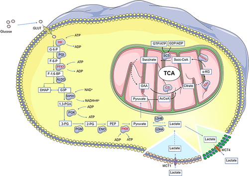
Figure 3 Schematic of lactate shuttling from cell to cell through the MCTs. Hypoxic cancer cells and cancer-associated fibroblasts (CAFs) use glucose to produce lactate through glycolysis, and lactate is transported out of the cell by MCT4. Aerobic cancer cells transport lactate through MCT1. Vascular endothelial cells transport lactic acid into cells through MCT1 and promote angiogenesis through NF-KB/IL8 pathway (㊉: activate).
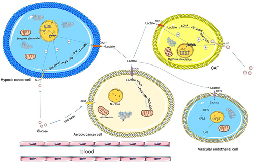
Figure 4 MCTs partial adjustment mode diagram.
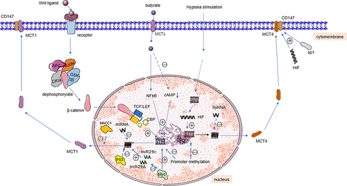
Table 1 Summary of MCTs Regulation
Table 2 Summary of Inhibitors