Figures & data
Figure 1 (A) shows a diffusely swollen left breast with shiny skin (blue arrow). (B and C) are ultra-sonographic images of the breast showing multiple large hypoechoic masses (blue stars) (4.95cm by 4.96cm and 5.82 cm) respectively, regularly shaped with no calcifications. (D) shows a touch imprint with medium-sized cells, coarse chromatin, and occasional tingible body macrophages (see black arrow).
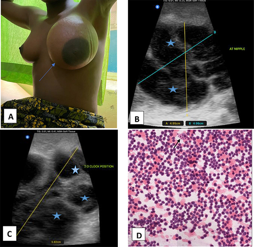
Figure 2 (A) (x20) shows an H&E stained section of the tumor with medium-sized cells, a high N: C ratio, and numerous tingible body macrophages (black arrows) giving it a characteristic starry sky. (B) shows a reduction in the size of the breast a week after her first cycle of chemotherapy with striae seen on the upper border of the breast (black arrowhead).
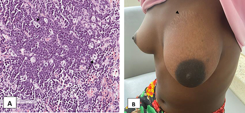
Figure 3 IHC stains showing positive CD45, Pax5, and c-MYC ((A–C) respectively) (x20). (D) shows 100% expression of Ki67. Panels E and F show negative Tdt and CD3 respectively.
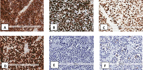
Figure 4 Left breast is now almost equal in size to the right breast during the third cycle of chemotherapy.