Figures & data
Table 1 Primer sequences used in reverse transcriptase polymerase chain reaction (RT-PCR)
Figure 1 The cytotoxicity of kefir on CEM and Jurkat cells.
Note: Error bars represent the standard deviation.
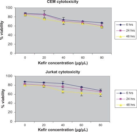
Figure 2 Effect of kefir on the viability of normal lymphocytes from the blood of healthy patients.
Notes: Error bars represent standard deviation. Kefir does not result in significant reduction in the viability of normal lymphocytes.
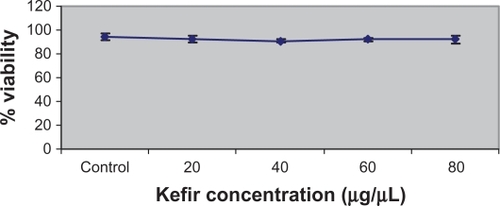
Figure 3 Effect of kefir on the proliferation of CEM and Jurkat cells.
Notes: The error bars represent standard deviation. Kefir causes a dose and time-dependent reduction in proliferation.
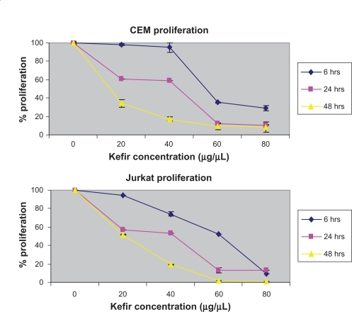
Figure 4 Effect of kefir on the cell cycle/apoptosis by flow cytometry on CEM and Jurkat cells. Results indicate that kefir causes a G0/G1 arrest, as evidenced by the increase in the cells in the pre-G1 phase.
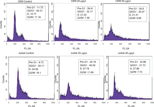
Figure 5 Effect of kefir on apoptosis using Cell Death ELISA kit, which quantitatively detects cytosolic histone-associated DNA fragments. Kefir causes an increase in DNA fragments, indicating an increase in apoptosis in both cell lines used.
Abbreviation: ELISA, enzyme-linked immunosorbent assay.
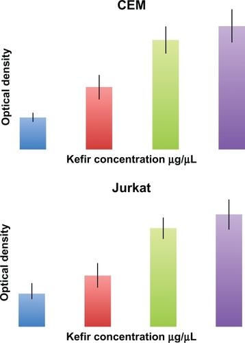
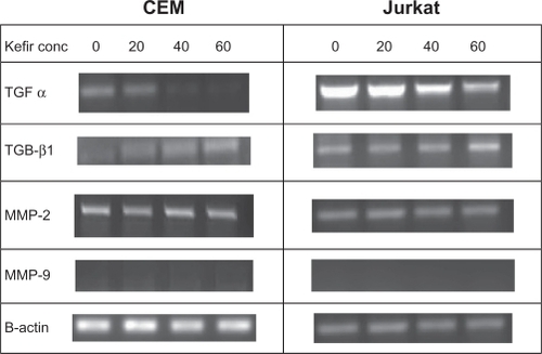
Figure 6 The effect of kefir on the transcriptional level of TGF-α, TGF β1, MMP-2 and MMP-9 in CEM and Jurkat cells.
Abbreviations: Conc, concentration; MMP, matrix metalloproteinase; TGF-α, trans forming growth factor-alpha; TGF-β1, transforming growth factor-beta1.