Figures & data
Figure 1 Initiator phase of geometric triangular chiral hexagonal like crystal complex assembly in malignant tumors. A and B show a geometric triangular chiral hexagonal-like crystal complex assembly in ascitic fluid in a case of abdominal carcinoma with Papanicolaou staining (40×); C and D show the triangular mirror image assembly in a case of endocervical adenocarcinoma, endocervical smear with Papanicolaou staining (40×, observe the black frame in A–C); E–G illustrate the crystal comet effect tail from the core of the hexagonal pattern in a case of cervical carcinoma in situ, vaginal smear with Papanicolaou staining (40×); H and I show the crystal comet effect tail with a simultaneously opposite crystal trajectory in a case of papillary thyroid carcinoma, fine needle aspiration, Papanicolaou staining (40×).
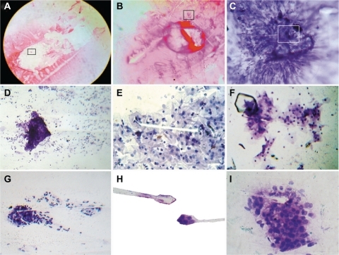
Table 1 Relationship between geometric triangular chiral crystal complexes and crystal comet effect tail assembly
Figure 2 The crystal comet effect tail generates triangular mirror images and a state-of-the-art complex. A shows the detachment subimage of B, which illustrates the triangular mirror image generated from the crystal comet effect tail in a case of endometrial adenocarcinoma, Papanicolaou staining (40×, observe the detail area inside the black frame); C shows the detachment subimage of D, which is a state-of-the-art complex in a case of mucinous cystadenocarcinoma of the ovary, ascitic fluid, Papanicolaou staining (40×); E is a detachment subimage of F, which shows embryoid body self-assembly into a geometric triangular chiral hexagonal like crystal complex in a case of colon adenocarcinoma with hematoxylin and eosin staining (20×); G is a detachment subimage of H, which shows fractal embryoid bodies ejected from the core of the hexagonal geometric complex, generating buoyant vibrational echo image waves in a case of breast adenocarcinoma, fine needle aspiration, Papanicolaou staining (20×); and I shows well-defined fractal embryoid bodies in a case of breast adenocarcinoma with hematoxylin and eosin staining (20×).
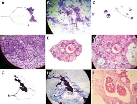
Table 2 Relationship between crystal comet effect tail assembly and embryoid body pattern
Figure 3 The crystal comet effect tail generates alignment of fractal embryoid bodies. A shows a detachment subimage of B, demonstrating fractal embryoid body alignment emerging from a hexagonal core geometric organization in a case of Grade 1 cervical intraepithelial neoplasia with Papanicolaou staining (10×); C is a detachment subimage of D which is a micrograph displaying assembly of well-defined fractal embryoid bodies in a case of gastric adenocarcinoma with hematoxylin and eosin staining (20×); E is a micrograph displaying assembly of well-defined fractal embryoid bodies in a case of colon adenocarcinoma with hematoxylin and eosin staining (20×); F is a detachment subimage of G, which shows a mirror image of well-defined fractal embryoid bodies in a case of renal cell carcinoma with hematoxylin and eosin staining (20×); H shows a fractal embryoid body in a case of undifferentiated sarcoma tumor with hematoxylin and eosin staining (20×); I shows self-assembled embryoid body with a well-defined pattern of formation in a necrotic area of leiomyosarcoma, with hematoxylin and eosin staining (20×).
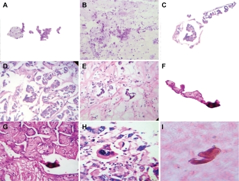
Figure 4 Well-defined self-assembled embryoid body pattern formation. A–D are closeup images of a well-defined embryoid body pattern formation in a case of squamous cell carcinoma of the skin, hematoxylin and eosin staining (20×); E–G show a well-defined, self-assembled embryoid body pattern formation in a case of peritoneal carcinoma with ascites, Papanicolaou staining (20×); H is a negative image of G; I reveals embryoid body self-assembly with geometric triangular chiral hexagonal crystal as a template platform in a case of peritoneal carcinoma with ascites and Papanicolaou staining (20×).
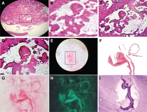
Figure 5 High-gradient self-assembled sequential embryoid body pattern formation. A is a detachment subimage of B, which shows high-gradient sequential embryoid body pattern formation from a case of breast adenocarcinoma, hematoxylin and eosin staining (20×); C shows a sequential embryoid body pattern with well-defined formation from a necrotic area in an undifferentiated sarcoma tissue, hematoxylin and eosin staining (20×); D is a negative image of C; E–G are closeup views of a well-defined embryoid body pattern formation in a case of breast adenocarcinoma, hematoxylin and eosin staining (20×); H is a negative image of G; I shows a well-defined pattern of embryoid body formation in a case of prostate adenocarcinoma, hematoxylin and eosin staining (20×).
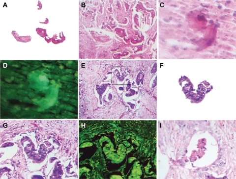
Figure 6 Macroscopic representation of an embryoid body. A is a macroscopic representation of a fractal embryoid body in a case of malignant ovarian tumor; B is a detachment subimage of C, a macroscopic representation of an embryoid body revealing a hexagonal geometric pattern and a crystal comet tail from a case of retroperitoneal malignant tumor; D is a detachment subimage of E, which is a macroscopic embryoid body representation from a case of renal cell carcinoma; F and G are macroscopic representations of fractal embryoid body structures from a case of leiomyosarcoma; H is a macroscopic representation of an embryoid body from a case of diffuse gastric cancer; I shows is an embryoid body from a case of squamous cell carcinoma of the skin, hematoxylin and eosin staining.
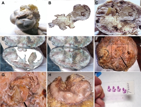
Figure 7 Neuron-specific enolase immunostaining analysis. A shows neuron-specific enolase immunostaining of control tissue from a peripheral nerve (10×); B reveals highly selective enolase immunopositivity in aligned fractal embryoid body structures from a case of esophageal squamous cell carcinoma (40×); C is a panoramic view revealing highly selective enolase immunopositivity in a self-assembled fractal embryoid body structure aligned into a hexagonal geometric pattern from a case of skin squamous cell carcinoma (10×); D is a detachment subimage of E, which shows highly selective enolase immunopositivity in a self-assembled embryoid body structure inside a hexagonal geometric pattern in a case of lung carcinoma (10×); F is a micrograph demonstrating highly selective enolase immunopositivity in a well-defined self-assembled embryoid body pattern in a case of undifferentiated lung cancer (20×); G is a micrograph revealing high enolase immunopositivity in a well-defined self-assembled embryoid body pattern from a case of prostate carcinoma (40×); H is a micrograph revealing enolase immunopositivity in a well-defined, self-assembled embryoid body pattern in a case of squamous cell carcinoma of the skin (10×); and I is a negative image of H.
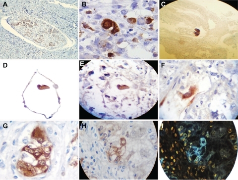
Table 3 Embryoid body selective distribution of neuron-specific enolase antibody immunopositivity
Figure 8 Comparative image analysis of reproducible and predictable template platforms generated by the electro-optical collision model. A shows a reproducible and predictable template platform of a comet tail, generated by the electro-optical collision model; B is a comparative image analysis of ascitic fluid from a case of ovarian carcinoma, with Papanicolaou staining (10×); C shows a reproducible and predictable template platform for geometric triangular chiral hexagonal-like crystal complexes generated by the electro-optical collision model; D is a comparative image analysis of a vaginal smear for a case of cervical carcinoma in situ, Papanicolaou staining (20×); E shows a reproducible and predictable template platform plasma magnetic circuit pattern generated by electro-optical collision; F comparative image analysis of a case of breast carcinoma, hematoxylin and eosin staining (20×); G shows well-defined pattern formation of an embryoid body in a case of colon adenocarcinoma, hematoxylin and eosin staining (20×), with comparative image analysis and a reproducible and predictable template platform plasma magnetic circuit pattern generated by electro-optical collision in H, which again shows a reproducible and predictable template platform plasma magnetic circuit pattern generated by electro-optical collision; I shows a welldefined embryoid body pattern formation in a case of colon adenocarcinoma, hematoxylin and eosin staining (20×), and comparative image analysis with a reproducible and predictable template platform plasma magnetic circuit pattern generated by electro-optical collision in H.
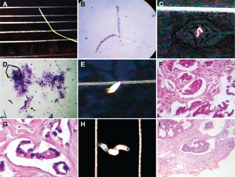
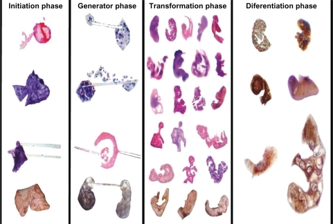
Figure 9 Sequential development of embryoid body pattern.