Figures & data
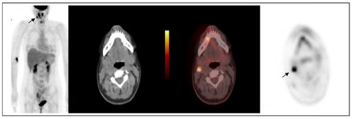
Figure 1 Fluorine-18 fluorodeoxyglucose PET/CT MIP and cross sectional images at the head and neck region and transthoracic level.
Notes: (A) The first PET/CT was carried out to rule out lymphoproliferative disease, it showed multiple small lesions in right lateral cervical and supraclavicular regions. It showed high 18-fluorodeoxyglucose uptake on the right side of the nasopharynx and a biopsy confirmed lymphoid hyperplasia of the palatine tonsil. (B) Two lesions in the right lung field. They are likely to represent an infectious foci.
Abbreviation: PET/CT, positron emission tomography/computed tomography.
Abbreviation: PET/CT, positron emission tomography/computed tomography.

Figure 2 18F-FDG PET/CT at 2 month intervals show reduction in size and number of cervical nodes (arrow) and that lesions in the lung field had disappeared.
Abbreviation: PET/CT, positron emission tomography/computed tomography.