Figures & data
Figure 1 A 45-year-old male smoker with pathologically diagnosed RB.
Abbreviations: CT, computed tomography; RB, respiratory bronchiolitis.
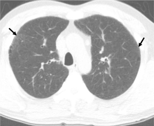
Table 1 Summary of key features
Figure 2 A 57-year-old male smoker with pathologically diagnosed DIP.
Notes: The axial CT image shows GGO in a subpleural and lower distribution. Thin-walled cysts (black arrows) are seen within the region of the GGOs. Image courtesy of Samsung Medical Center.
Abbreviations: CT, computed tomography; DIP, desquamative interstitial pneumonia; GGO, ground-glass opacity.
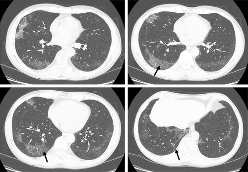
Figure 3 CT features of AEF.
Abbreviations: AEF, airspace enlargement with fibrosis; CT, computed tomography.
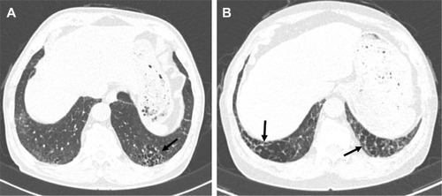
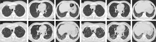
Figure 4 A 26-year-old male ex-smoker with pathologically diagnosed RB.
Abbreviations: CT, computed tomography; RB, respiratory bronchiolitis.
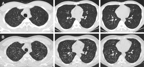
Figure 5 A 66-year-old male ex-smoker with lung biopsy-proven RB-ILD.
Notes: There were bronchiolocentric GGOs and traction bronchiectasis in both lungs that represented airway-centered interstitial fibrosis. Image courtesy of Samsung Medical Center.
Abbreviations: GGO, ground-glass opacity; ILD, interstitial lung disease; RB, respiratory bronchiolitis.
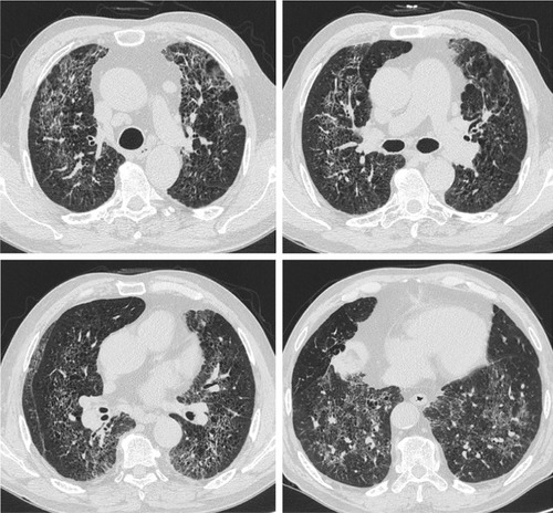
Figure 6 A 44-year-old male smoker with pathologically diagnosed RB-ILD.
Abbreviations: CT, computed tomography; GGO, ground-glass opacity; ILD, interstitial lung disease; RB, respiratory bronchiolitis; UIP, usual interstitial pneumonia.