Figures & data
Figure 1 Schematic diagram of the position of the multipair esophageal electrode and surface electrode.
Abbreviations: EMGdi,es, transesophageal diaphragmatic electromyogram activity; EMGdi,sur, surface diaphragmatic electromyogram activity; EMGpara, surface electromyogram activity of the parasternal intercostal muscles; EMGsc, surface electromyogram activity of the sternocleidomastoid muscles.
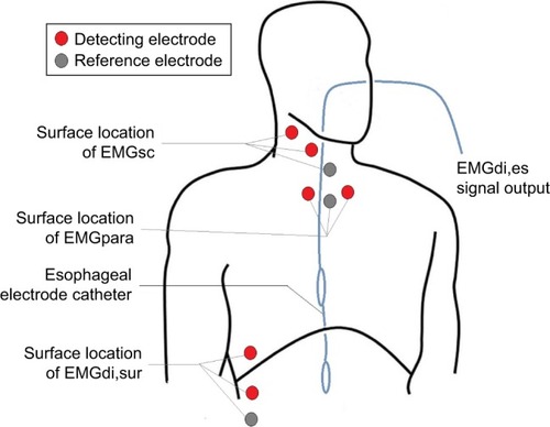
Table 1 Variance of transesophageal diaphragmatic electromyogram activity (EMGdi,es) and surface inspiratory electromyogram activity during treadmill exercise
Figure 2 Waveform of esophageal pressure, transesophageal diaphragmatic electromyogram activity, and surface inspiratory electromyogram activity.
Abbreviations: EMGdi1, first transesophageal diaphragmatic electromyogram; EMGdi2, second transesophageal diaphragmatic electromyogram; EMGdi3, third transesophageal diaphragmatic electromyogram; EMGdi4, fourth transesophageal diaphragmatic electromyogram; EMGdi5, fifth transesophageal diaphragmatic electromyogram; EMGdi,sur, surface diaphragmatic electromyogram activity; EMGpara, surface electromyogram activity of the parasternal intercostal muscles; EMGsc, surface electromyogram activity of the sternocleidomastoid muscles; Pes, esophageal pressure.
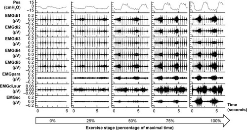
Figure 3 Changing characteristics of RMSdi,es and surface inspiratory electromyogram activity during increased treadmill exercise capacity.
Abbreviations: RMS, root mean square; RMSdi,es, RMS of transesophageal diaphragmatic electromyogram activity; RMSdi,sur, RMS of the surface electromyogram activity of the diaphragm; RMSpara, RMS of the surface electromyogram activity of the parasternal intercostal muscles; RMSsc, RMS of the surface electromyogram activity of the sternocleidomastoid muscles.
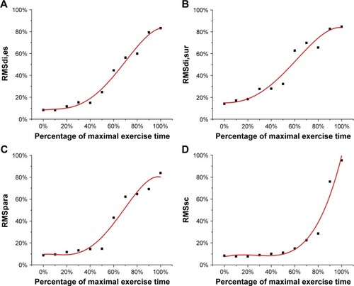
Figure 4 Scatter plot for correlation between RMSdi,es and RMS of the surface inspiratory muscles EMG.
Abbreviations: RMS, root mean square; EMG, electromyogram; RMSdi,es, RMS of transesophageal diaphragmatic electromyogram activity; RMSdi,sur, RMS of the surface electromyogram activity of the diaphragm; RMSpara, RMS of the surface electromyogram activity of the parasternal intercostal muscles; RMSsc, RMS of the surface electromyogram activity of the sternocleidomastoid muscles.
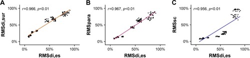
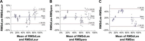
Figure 5 Bland-Altman plots for reliability of estimation between RMSdi,es and RMS of surface inspiratory muscles EMG.
Abbreviations: RMS, root mean square; EMG, electromyogram; RMSdi,es, transesophageal diaphragmatic electromyogram activity; RMSdi,sur, RMS of the surface electromyogram activity of the diaphragm; RMSpara, RMS of the surface electromyogram activity of the parasternal intercostal muscles; RMSsc, RMS of the surface electromyogram activity of the sternocleidomastoid muscles.