Figures & data
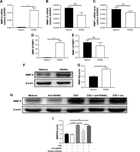
Figure 1 Mouse lung function and pulmonary emphysema.
Notes: (A) TLC and (B) airway resistance (R) were measured in mice; n=6. Lung histology of mice that were exposed to (C) CS and (D) air, by HE staining (400×), scale bar=50 µm. (E) MLI and (F) DI were measured in mice; n=6. Bars indicate the mean value and error bars indicate SEM. *P<0.05 and ***P<0.001.
Abbreviations: CS, cigarette smoke; DI, destructive index; MLI, Mean linear intercept; SEM, standard error of the mean; TLC, Total lung capacity.
Abbreviations: CS, cigarette smoke; DI, destructive index; MLI, Mean linear intercept; SEM, standard error of the mean; TLC, Total lung capacity.
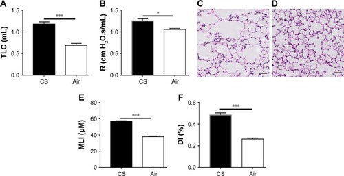
Figure 2 Expression and cellular localization of RANKL and RANK in CS-exposed mice.
Notes: Immunofluorescent staining for RANKL in (A) CS-exposed mice and (C) air-exposed mice. Immunofluorescent staining for RANK in (B) CS-exposed mice and (D) air-exposed mice. (E) Co-immunofluorescent staining for RANKL (AlexaFluor 594, red) and F4/80 (AlexaFluor 488, green) in the lungs of CS-exposed mice. (F) Co-immunofluorescent staining for RANK (AlexaFluor 594, red) and F4/80 (AlexaFluor 488, green) in the lungs of CS-exposed mice. Sections were counterstained with DAPI (blue). Arrows indicate positive cells. (A, B, E and F) Scale bar=25 µm. (C and D) Scale bar=50 µm.
Abbreviations: CS, cigarette smoke; RANKL, receptor activator of nuclear factor-κB ligand.
Abbreviations: CS, cigarette smoke; RANKL, receptor activator of nuclear factor-κB ligand.
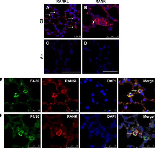
Figure 3 CSE promotes RANKL and RANK expression in vitro.
Notes: MH-S and RAW264.7 cell lines were stimulated as in medium and with 0.5% CSE, and RANKL expression was detected by flow cytometry. The MFI of RANKL measured in (A and C) MH-S and (B and D) RAW264.7 cells. MH-S and RAW264.7 cells were stimulated as in medium and with 0.5% CSE, and RANK expression was detected by flow cytometry. The MFI of RANK measured in (E and G) MH-S and (F and H) RAW264.7 cells. Black: medium stained with isotype antibody-phycoerythrin; red: medium; green and blue: CSE. (C, G and H) n=3; (D) n=5. Bars indicate the mean value, and error bars indicate SEM. *P<0.05, **P<0.01 and ***P<0.001.
Abbreviations: CSE, cigarette smoke extract; MFI, mean fluorescence intensity; PE, phycoerythrin; RANKL, receptor activator of nuclear factor-κB ligand; SEM, standard error of the mean.
Abbreviations: CSE, cigarette smoke extract; MFI, mean fluorescence intensity; PE, phycoerythrin; RANKL, receptor activator of nuclear factor-κB ligand; SEM, standard error of the mean.
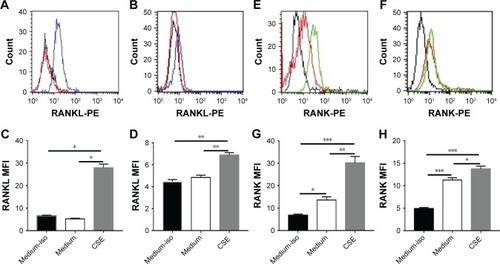
Figure 4 Alveolar macrophages under CSE stimulation have reduced RANK protein degradation and translocation to the lysosomal compartment.
Notes: Co-immunofluorescent staining for LAMP1 (AlexaFluor 488, green) and RANK (AlexaFluor 594, red) in (A) unstimulated macrophages and (B) CSE stimulated macrophages for 24 hours; nuclei were stained with DAPI (blue). (C and D) Representative Western blot and relative quantification normalized to β-actin for Rab7 in MH-S stimulated with CSE for 24 hours; n=3. Bars indicate the mean value, and error bars indicate SEM. *P<0.05.
Abbreviations: CSE, cigarette smoke extract; RANKL, receptor activator of nuclear factor-κB ligand; SEM, standard error of the mean.
Abbreviations: CSE, cigarette smoke extract; RANKL, receptor activator of nuclear factor-κB ligand; SEM, standard error of the mean.
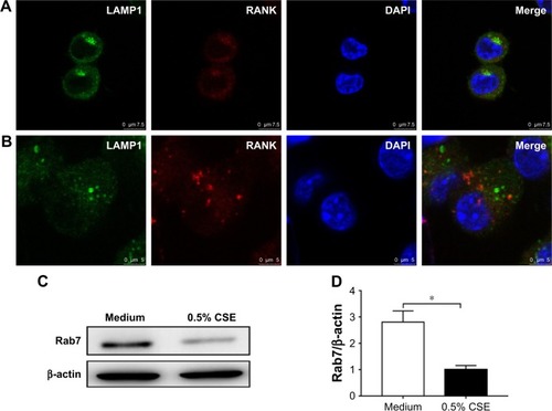
Figure 5 Upregulation of MMP-9 expression by CS.
Notes: Immunohistochemical detection of MMP-9 (DAB, brown) in (A) CS-exposed mice and (B) air-exposed mice. Sections were counterstained with Mayer hematoxylin (blue). Scale bar=50 µm. (C) Number of MMP-9–positive cells per square millimeter, n=5. (D) MMP-9 mRNA level in the lung tissues, n=4. (E) Co-immunofluorescent staining for MMP-9 (AlexaFluor 594, red) and F4/80 (AlexaFluor 488, green) in the lungs of CS-exposed mice. Nuclei were stained with DAPI (blue). (F and G) Representative Western blot and relative quantification normalized to β-actin for MMP-9 in MH-S stimulated with CSE for 48 hours; n=3. Arrows indicate positive cells. Bars indicate the mean value, and error bars indicate SEM. *P<0.05, **P<0.01 and ***P<0.001.
Abbreviations: CS, cigarette smoke; CSE, CS extract; SEM, standard error of the mean.
Abbreviations: CS, cigarette smoke; CSE, CS extract; SEM, standard error of the mean.
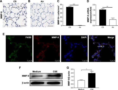
Figure 6 MMP-9 expression was regulated by RANKL.
Notes: (A) MMP-9 mRNA was upregulated in MH-S under RANKL stimulation. (B and C) MMP-12 and TIMP1 mRNA in MH-S under RANKL stimulation. The ratio of (D) MMP-9 mRNA to TIMP1 mRNA and (E) MMP-12 mRNA to TIMP1 mRNA under RANKL stimulation. (F and G) Representative Western blot and relative quantification normalized to β-actin for MMP-9 in MH-S stimulated with RANKL. (H and I) Representative Western blot and relative quantification normalized to β-actin for MMP-9 in MH-S stimulated with combined CSE with monoclonal anti-RANKL antibody. n=3. Bars indicate the mean value, and error bars indicate SEM. *P<0.05, **P<0.01 and ***P<0.001.
Abbreviations: CSE, cigarette smoke extract; NS, not significant; iso, isotype antibody; SEM, standard error of the mean.
Abbreviations: CSE, cigarette smoke extract; NS, not significant; iso, isotype antibody; SEM, standard error of the mean.