Figures & data
Table 1 Primer sets for RT-PCR analysis assay
Figure 1 PM exposure induces SIRT1 reduction in HBE cells. (A) Cells were exposed to PM at indicated times or concentrations. Western blot analysis for SIRT1 protein expression in HBE cells treated with PM. (B) Immunofluorescence for SIRT1 protein expression in HBE cells treated with PM (Magnification×400). (C) Semi-quantification results of immunofluorescence. (D and E) RT-PCR analysis for a time course (PM at 100 μg/mL for various times) or dose response (various concentrations of PM for 24 h) of SIRT1 protein expression in HBE cells treated with PM. Data are presented as mean ± SD of three independent experiments. *P<0.05, **P<0.01, and ***P<0.001.
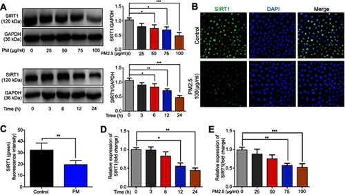
Figure 2 SIRT1 gene silence increases PM-induced inflammatory cytokines in HBE cells. Cells were transfected with control (CTL) siRNA or SIRT1 siRNA for 24 h, and then were treated with PM (100 μg/mL) for 24 h to measure the levels of IL-6, IL-8, IL-17A, and MUC5AC.(A) SIRT1 expression was measured by Western blot. (B-F) The mRNA levels of IL-6, IL-8, IL-17A, and MUC5AC were measured by RT-PCR. Data are presented as mean ± SD of three independent experiments. **P<0.01 and ***P<0.001.
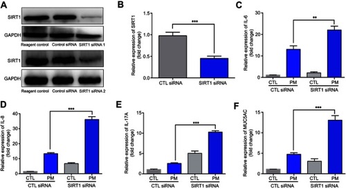
Figure 3 Resveratrol (Res) inhibits PM-induced inflammatory cytokines in HBE cells. HBE cells were treated with SIRT1 activator resveratrol (Res) together with PM for 24 h. (A-E) The mRNA levels of IL-6, IL-8, IL-17A, and MUC5AC were measured by RT-PCR. Data are presented as Mean ± SD of three independent experiments. *p<0.05, **p<0.01, ***p<0.001, and ****p<0.0001.
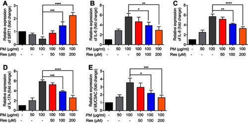
Figure 4 Resveratrol (Res) inhibits PM-induced airway inflammation in mice. Mice were instilled intratracheally with normal saline (NS), PM or PM plus resveratrol (Res) for 3 d, and the mice were sacrificed after 24 h. (A) Western blot analysis for SIRT1 protein expression in lung tissues of mice. (B) IHC analysis for representative images of lung tissues of mice stained with SIRT1 protein (red arrows; Magnification×400) and semi-quantified SIRT1 expression of the IHC staining. (C) The total inflammatory cells and the number of neutrophils (D) in the BALF were measured. (E) Representative images of macrophages and neutrophils in the BALF were analyzed (red arrows; Magnification×400) using Giemsa staining. (F) Representative images of lung tissue stained with H&E (Magnification×400) and Semi-quantified inflammation score of the H&E staining (red arrows). Data are representative of three independent studies (n=5–6 mice per group per study). Results are expressed as mean ± SD. *p<0.05, **p<0.01, ***p<0.001, and ****p<0.0001.
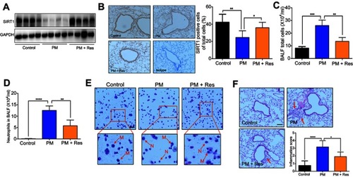
Figure 5 Resveratrol (Res) reduces airway inflammation in response to PM exposure. The levels of IL-6, IL-8, IL-17A, TNF-α, and MUC5AC in lung tissue were analyzed by RT-PCR. Data are representative of three independent studies (n=5–6 mice per group per study). Results are expressed as mean ± SD. *p<0.05, **p<0.01, and ***p<0.001. Abbreviations: PM, Particulate matter; MUC5AC, Mucin 5AC; RT-PCR, Reverse Transcription-Polymerase Chain Reaction.
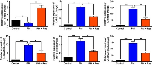
Figure 6 Resveratrol (Res) suppresses PM-induced inflammation response in HBE cells and mice via MAPK pathways. (A) HBE cells were treated with PM or resveratrol (Res), and the protein levels of MAPK pathways were measured by Western blot. (B) Mice were instilled intratracheally with normal saline (NS), PM or PM plus resveratrol respectively for 3 days, and the mice were sacrificed after 24 h. The protein levels of MAPK pathways were measured by Western blot. Data are representative of three independent studies. Data are representative of three independent studies. Results are expressed as mean ± SD. *p<0.05.
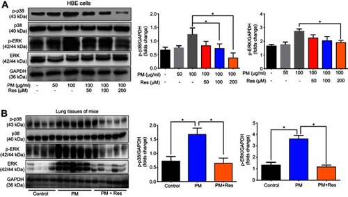
Figure 7 MAPK inhibitors suppress PM-induced inflammation response in HBE cells. (A–I) p38 inhibitor SB203580 and ERK inhibitor U0126 were added 2 h prior PM exposed and cells were collected after 24 h. The levels of TNF-α, IL-6 and, IL-17A were analyzed by RT-PCR. Data are representative of three independent studies. Results are expressed as mean ± SD. *p<0.05, **p<0.01, ***p<0.001, and ****p<0.0001.
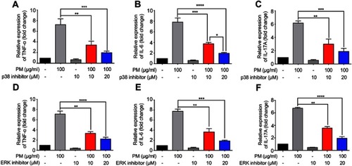
Figure S1 SIRT1 overexpression suppresses PM-induced inflammation response in HBE cells. Cells were transfected with Control or SIRT1 plasmid and were stimulated with PM. (A) SIRT1 expression was measured by Western blot. (B-F) The mRNA levels of SIRT1, IL-6, IL-8, IL-17A, and MUC5AC were assessed by RT-PCR.Data are representative of three independent experiments. Results are expressed as mean ± SD. *p<0.05, **p<0.01, and ****p<0.0001.
Abbreviations: SIRT1, Sirtuin 1; PM, Particulate matter; HBE, Human bronchial epithelial.
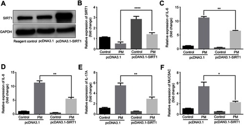
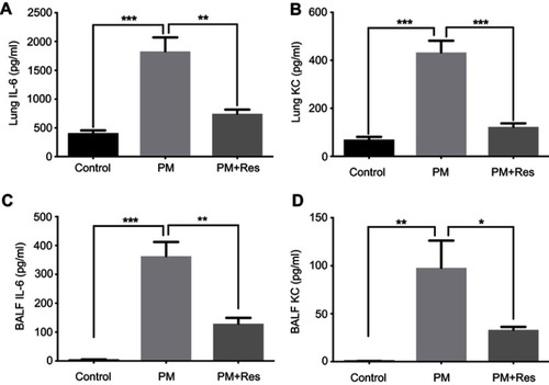
Figure S2 Resveratrol (Res) suppresses PM-induced airway inflammation. WT mice were exposed to PM for 3 days. Lungs and BALF were isolated 1 day after the last PM challenge. The inflammatory cytokines in the lung homogenate (A and B) and BALF (C and D) were assessed by using an enzyme-linked immunosorbent assay. Data are representative of three independent studies (n=5–6 mice per group per study). Results are expressed as mean ± SD. *p<0.05, **p<0.01, and ***p<0.001.
Abbreviations: PM, Particulate matter; WT, Wild type; BALF, Brochoalveolar lavage fluid; KC, Keratinocyte-derived Cytokine.