Figures & data
Figure 1 MHFM of VTF in COPD.
Notes: (A) Representative MAG and PC images from the proximal (red contour) and distal (green contour) portions of the RPA. (B) Representative mean velocity profiles over the cardiac cycle measured in the proximal (red trace) and distal (green trace) portions of the RPA. (C) Representative examples of Fourier transform magnitudes and VTF in subjects with high (>1) and low (<1) MHFM (average modulus for harmonics 5–7).
Abbreviations: MHFM, mean high-frequency modulus; VTF, velocity transfer function; MAG, magnitude; PC, phase-contrast; RPA, right pulmonary artery.
Abbreviations: MHFM, mean high-frequency modulus; VTF, velocity transfer function; MAG, magnitude; PC, phase-contrast; RPA, right pulmonary artery.
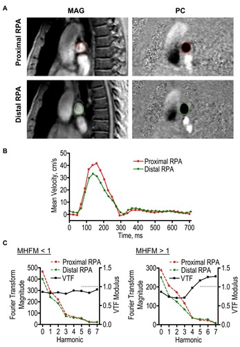
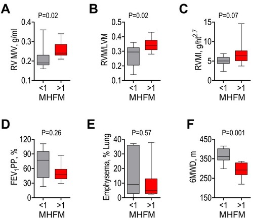
Figure 2 Cardiac and pulmonary measurements in COPD with high (>1) and low (<1) MHFM of VTF.
Notes: High and low MHFM groups had different RVM/V (A), RVM/LVM (B); but did not differ by RVMI (C), FEV1 PP (D), or emphysema (E). However, 6MWD was lower in the high MHFM group (F).
Abbreviations: MHFM, mean high-frequency modulus; VTF, velocity transfer function; RVM/V, right ventricular mass to volume ratio; RVM/LVM, ratio of right and left ventricular mass; RVMI, right ventricular mass index; FEV1 PP, percent predicted forced expiratory volume in 1-second; 6MWD, 6-mins walk distance.
Abbreviations: MHFM, mean high-frequency modulus; VTF, velocity transfer function; RVM/V, right ventricular mass to volume ratio; RVM/LVM, ratio of right and left ventricular mass; RVMI, right ventricular mass index; FEV1 PP, percent predicted forced expiratory volume in 1-second; 6MWD, 6-mins walk distance.