
Figures & data
Figure 1 Schematic depiction of chitin, chitinases and chitinase-like proteins (CLPs) involvement in lung diseases.
Notes: Exposure to chitin may trigger the secretion of chitinases and CLPs from neutrophils, macrophages and other cells (solid-line black arrows show cell activation after chitin exposure and secretion of chitinases). Both groups of enzymes are thought to play a role in the pathogenesis of pulmonary diseases (eg, asthma and COPD). Black dashed arrows reflect a negative (ie, destructive) effect, while the grey dashed arrows show a possible positive (ie, protective) influence. See manuscript text for more details.
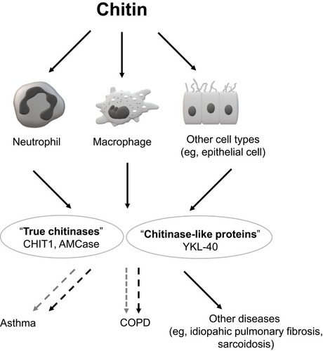
Figure 2 Schematic presentation of the hypothesized links between chitotriosidase (CHIT1) and the pathogenesis of lung inflammation and injury (eg, in COPD).
Notes: The exposure to cigarette smoke and other factors may enhance CHIT1 expression and increase its production, mainly by lung macrophages and neutrophils. The impact of CHIT1 on lung cells is heterogeneous and include inflammation and tissue destruction, mediated by interleukin 8 (IL-8), metalloproteinase 9 (MMP-9) and monocyte chemoattractant protein 1 (MCP-1), as well as lung fibrosis induced by transforming growth factor-beta (TGF-β) pathway. However, the exact role of CHIT1 in the pathogenesis of lung diseases is still highly hypothetical. A large white arrow represents stimulation of macrophages and neutrophils by various factors, the thick grey arrows show the secretion of CHIT1 from the cellular sources, the thin black arrows depict the pathways of CHIT1 activity, the vertical yellow arrow represents the increase or enhancement.
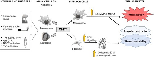
Figure 3 Schematic representation of the supposed mechanisms involved in YKL-40-related emphysematous lung destruction, tissue inflammation and remodeling.
Notes: The large white arrow represents stimulation of macrophages, epithelial cells and neutrophils by various factors, the thin dashed black arrow depicts an indirect effect of environmental factors, the thick grey arrows show the secretion of YKL-40 from the cellular sources, the thin black arrows depict the possible YKL-40 signaling pathways, the thin dashed grey arrow depicts a stimulatory effect of interleukin 13 (IL-13) on transforming growth factor-beta (TGFβ), the vertical yellow arrow represents the increase or enhancement. See manuscript text for more details.
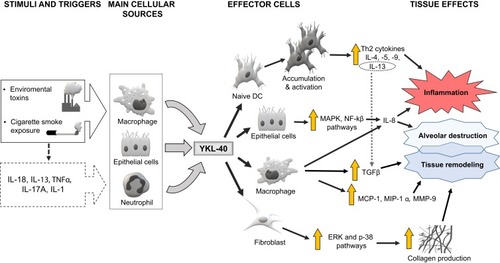
Table 1 Selected Information About Main Chitinases and Their Role in Humans