Figures & data
Figure 1 PRISMA flow chart. Studies were excluded both at screening and at full-text review, leading to a total of 51 studies included in the qualitative synthesis.
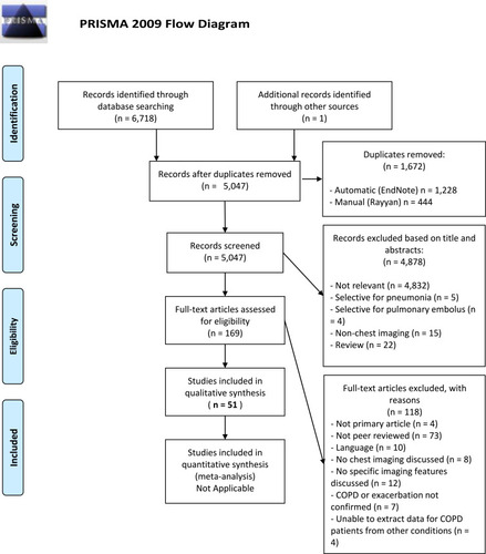
Table 1 Imaging Features (“Biomarkers”) at Exacerbation of COPD
Figure 2 Summary: The Pathophysiology of a COPD Exacerbation in relation to Imaging Biomarkers. An exacerbation of COPD is associated with an increase in airway wall area and airway resistance, a decrease in airway calibre, hyperinflation, an increase in the diameter of the main pulmonary artery, a decrease in the number of visible small pulmonary vessels and cardiac enlargement. These features are suggestive of additional inflammation and mucus in the lumen of the airways, increased obstruction in the small airways and air trapping, pulmonary hypertension and right-sided cardiac impairment.
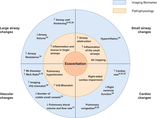
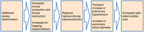
Figure 3 Hypothesis of the Pathophysiological Cascade Characteristic to an Exacerbation of COPD. Imaging features present during an exacerbation of COPD and their underlying pathophysiological processes motivate a hypothesis about the sequence of events in the lung which characterize an exacerbation. An exacerbation of COPD may be a cascade of events triggered by the onset of additional inflammation in the lumen of the airways, which leads to airway wall inflammation, bronchoconstriction and the production of excess mucus. This airway obstruction results in hyperinflation and the characteristic symptom of increased dyspnoea, and reactive vasoconstriction in small pulmonary vessels. In turn, this vasoconstriction leads to a transient increase in pulmonary arterial pressure, causing an enlargement of the pulmonary artery and right-sided cardiac dysfunction.