Figures & data
Figure 1 Representative photomicrograph of small airways disease and emphysema in a patient with chronic obstructive pulmonary disease.
Notes: The small airway (left panel) is characterized by obstruction of the lumen by mucous exudate. Also note the markedly thickened airway wall with recruitment of numerous inflammatory cells. In the right panel, emphysema is characterized by destruction of alveolar walls without enlargement of airspace, and without obvious fibrosis. Bar 30 μm.
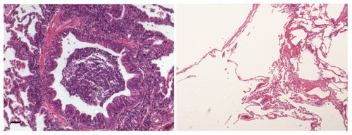
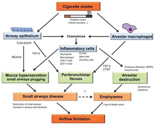
Figure 2 Diagram depicting potential mechanisms leading to airflow limitation via structural abnormalities in the small conducting airways and alveoli.
Notes: Cigarette smoke, a source of oxidative stress and irritants, is recognized by airway epithelial cells and alveolar macrophages. Secretion of chemokines by resident cells induces recruitment of inflammatory cells in the airways and alveoli.Citation33 Recognition of cigarette smoke by the airway epithelium involves TLR-4 and leads to the activation of EGFR, resulting in mucus production in epithelial goblet cells.Citation46 Neutrophil proteases (eg, elastase) activate the degranulation of goblet cells,Citation48 contributing to formation of mucous exudates within the lumen of the small airway. The repair response in small airways also involves thickening of the airway walls, at least in part related to peribronchial fibrosis. Activation of fibrosis in peribronchiolar mesenchymal cells is related to secretion of profibrotic growth factors (eg, TGF-β and CTGF) released by the airway epithelium and by alveolar macrophages.Citation12,Citation33 Small airways disease, characterized by an increase in airway wall thickness and mucous exudates in the lumen,Citation11 and by destruction of the small airways,Citation10 contributes to reduction in the cross-sectional area of the small airways, leading to airflow limitation. Alveolar destruction, which characterizes emphysema, may be related to proteolytic destruction of alveolar walls and/or to the failure of lung maintenance and repair programs involving apoptosis, senescence, and autoimmunity.Citation20 Emphysema promotes airway closure during expiration, contributing to airflow limitation via loss of elastic recoil. Recent data also suggest that destruction of the small airways may promote emphysema via loss of support at the distal acinus.Citation10,Citation30
Abbreviations: EGFR, epidermal growth factor receptor; TGF-β, transforming growth factor; TLR-4, toll-like receptor-4; CTGF, connective tissue growth factor; MMPs, matrix metalloproteinases.
Abbreviations: EGFR, epidermal growth factor receptor; TGF-β, transforming growth factor; TLR-4, toll-like receptor-4; CTGF, connective tissue growth factor; MMPs, matrix metalloproteinases.