Figures & data
Table 1 Three Common Bronchial Variation Subtypes and Clinical Abnormalities
Figure 1 Rate of decline in FEV1 with genetic and environmental factors. This downtrend of FEV1 over a lifetime is determined by genetics and epigenetics before birth. Then the different curves decline in different rates dependent on organ development/repair (aging) and environmental factors (smoking and air pollutions) after birth. The lung function of people with bronchial variations is worse than that of normal people at birth. The risk of COPD is higher and earlier if environmental exposure is not taken into account.
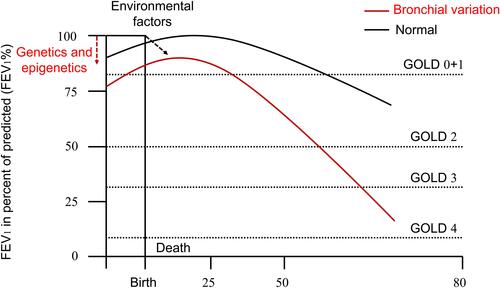
Figure 2 A model for the opposing effect of Fgf10 and Bmp4 in lung bud morphogenesis. An extending bud is shown schematically. Purple and yellow denote Bmp4 and Fgf10 expression. (A) Throughout bronchial development, Bmp4 is expressed in the epithelium and Fgf10 in the adjacent mesenchyme. Fgf10 promotes lung endoderm proliferation and migration. Bmp4 antagonizes Fgf10-mediated outgrowth of lung endoderm. The balance between the expression of Fgf10 and Bmp4 exists during branching, keeping the normal development of bronchus. (B) As the expression of Fgf10 increased, distal mesenchyme has shown hypertrophic morphologically and inhibited the expression of Bmp4 in the epithelium to stop branching. (C) Restricted by the traction of the internal epithelium, the expression of Fgf10 on both sides increased gradually, and lateral buds appeared symmetrically which represented as bifurcating. (D) Once the lung buds have been initiated, the expression of Bmp4 increased, and the expression of Fgf10 was inhibited. Epithelium grew along the distal mesenchyme towards lateral buds. The cycle of promotion of mesenchymal proliferation and outgrowth of endoderm begins again. (E) We considered the alternative hypothesis that the expression of Fgf10 at the tip of one lung bud increased excessively and downregulated Bmp4 expression. Then this bud formed a blind end or diverticulum with only another bud growing which was called absent variation (eg RMB or RLB in bridging bronchus). (F) When Bmp4 was more predominantly expressed than Fgf10, endothelial growth was faster than mesenchyme differentiation. Then more than three buds appeared at the bifurcation which was called accessory variation (eg tracheal bronchus, accessory cardiac bronchus).
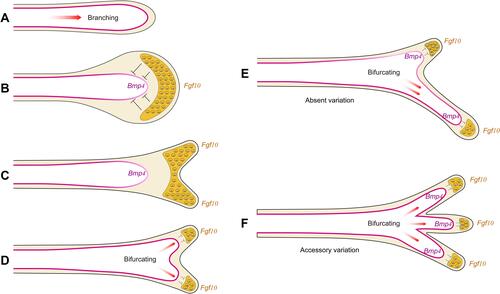
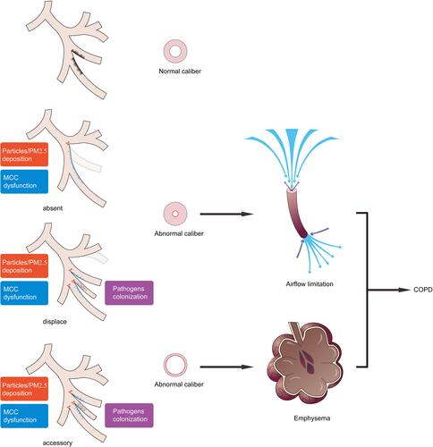
Figure 3 Schematic representation of bronchial variations in the development of COPD. Bronchial variations participate in the pathogenesis of COPD as anatomical determinants and promote abnormal bifurcations with more particles deposition. Abnormal variant bronchus covered cilia with movement and distribution dysfunction (MCC dysfunction) allowed more pathogens colonization. Absent or displaced bronchial variation presents bronchial wall thickening and caliber shorter, resulting in airflow limitation. Accessory or displaced bronchial variation accompanied by bronchial wall thinning and lumen enlargement results in emphysema. These are two common pathophysiological development patterns of COPD.