Figures & data
Table 1 Characteristics of Subjects in This Study
Figure 1 CircANKRD11 was overexpressed in the tissues of COPD patients and CSE-induced HPMECs. (A) CircANKRD11 expression was detected by qRT-PCR in the lung tissues of non-smokers (N=10), smokers without COPD (N=17) and smokers with COPD (N=21). (B) CircANKRD11 expression was determined by qRT-PCR in HPMECs after treatment of CSE (0%, 0.5%, 1.0%, 2.5% and 5%) for 24 h. (C) 2.5% CSE was exposed into HPMECs for 0, 6, 12, 24 and 36 h, respectively, and circANKRD11 expression was determined by qRT-PCR. *P<0.05.
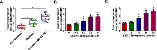
Figure 2 CircANKRD11 absence protected HPMECs from CSE-induced injury. (A–I) HPMECs were treated with 0% CSE (Control), 2.5% CSE, 2.5% CSE+si-NC and 2.5% CSE+si-circANKRD11, respectively. (A) CircANKRD11 expression was determined by qRT-PCR. (B) Cell apoptosis was detected by Annexin V-FITC and PI double staining assay. (C) The protein expression of Bcl-2, Bax, c-caspase 3 and p-caspase 3 was determined by Western blot analysis. (D–F) The levels of IL-1β, IL-6 and TNF-α were detected by ELISA. (G) The level of ROS was detected by ROS detection assay. (H) MDA level was detected by MDA determination assay. (I) SOD activity was detected by SOD activity detection assay. *P<0.05.
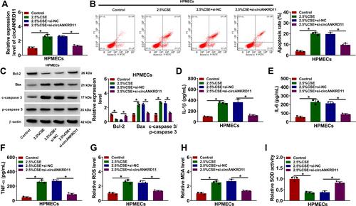
Figure 3 CircANKRD11 directly bound to miR-145-5p in HPMECs. (A) Circinteractome online database was employed to predict the binding sites between circANKRD11 and miR-145-5p. (B) MiR-145-5p expression was detected by qRT-PCR in HPMECs transfected with miR-NC, miR-145-5p, in-miR-NC or in-miR-145-5p. (C–E) Dual-luciferase reporter, RIP and RNA pull-down assays were employed to demonstrate the binding relationship between circANKRD11 and miR-145-5p in HPMECs. (F) MiR-145-5p expression was detected by qRT-PCR in the lung tissues of non-smokers (N=10), smokers without COPD (N=17) and smokers with COPD (N=21). (G) MiR-145-5p expression was determined by qRT-PCR in 2.5% CSE-treated HPMECs and untreated HPMECs. (H) The impacts between circANKRD11 silencing and miR-145-5p inhibitors on miR-145-5p expression were determined by qRT-PCR in HPMECs. *P<0.05.
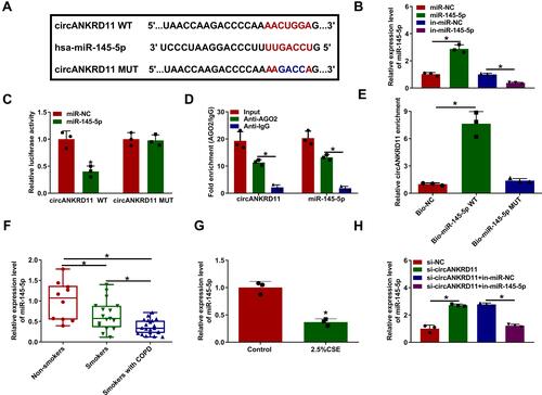
Figure 4 CircANKRD11 knockdown repressed cell apoptosis, inflammation, and oxidative stress by interacting with miR-145-5p in CSE-induced HPMECs. (A–H) 2.5% CSE-induced HPMECs were severally transfected with si-NC, si-circANKRD11, si-circANKRD11+in-miR-NC and si-circANKRD11+in-miR-145-5p. (A) Annexin V-FITC and PI double staining assay was employed to detect cell apoptosis. (B) The protein levels of Bcl-2, Bax, c-caspase 3 and p-caspase 3 were determined by Western blot analysis. (C–E) The levels of IL-1β, IL-6 and TNF-α were detected by ELISA. (F) The level of ROS was detected by ROS detection assay. (G) MDA level was detected by MDA determination assay. (H) SOD activity was detected by SOD activity detection assay. *P<0.05.
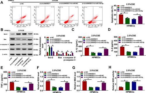
Figure 5 CircANKRD11 modulated BRD4 via binding to miR-145-5p. (A) The binding sites between miR-145-5p and BRD4 were predicted by microT_CDS online database. (B) Luciferase activities were detected by dual-luciferase reporter assay in HPMECs. (C and D) The mRNA and protein expression of BRD4 were determined by qRT-PCR and Western blot, respectively, in the lung tissues of non-smokers (N=10), smokers without COPD (N=17) and smokers with COPD (N=21). (E) BRD4 protein expression was detected by Western blot analysis in HPMECs treated with 0% CSE or 2.5% CSE. (F) The effects between miR-145-5p mimics and BRD4 overexpression on the protein expression of BRD4 were determined by Western blot analysis in HPMECs. (G) The impacts between circANKRD11 silencing and miR-145-5p inhibitors on BRD4 protein expression were revealed by Western blot analysis in HPMECs. *P<0.05.
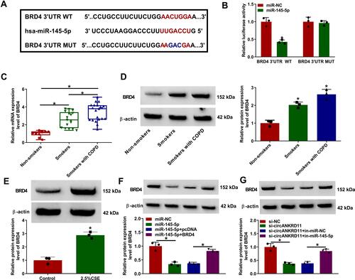
Figure 6 MiR-145-5p mimics protected against CSE-induced HPMEC injury through interacting with BRD4. (A–H) HPMECs were transfected with miR-NC, miR-145-5p, miR-145-5p+pcDNA and miR-145-5p+BRD4, respectively, under 2.5% CSE treatment in HPMECs. (A) Cell apoptosis was detected by Annexin V-FITC and PI double staining assay. (B) The protein expression of Bcl-2, Bax, c-caspase 3 and p-caspase 3 was determined by Western blot analysis. (C–E) The levels of IL-1β, IL-6 and TNF-α were detected by ELISA. (F) The level of ROS was detected by ROS detection assay. (G) MDA level was detected by MDA determination assay. (H) SOD activity was detected by SOD activity detection assay. *P<0.05.
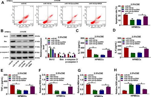
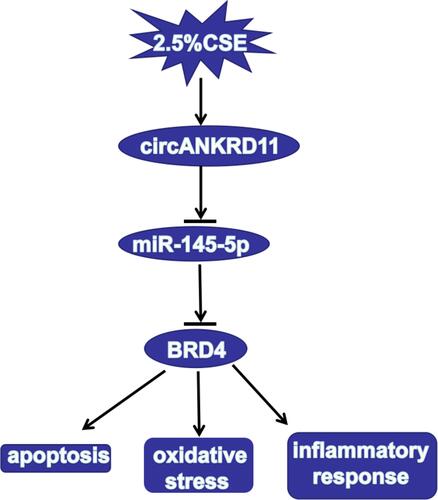
Figure 7 The illustration of circANKRD11 in regulating CSE-induced cell injury of HPMECs. CircANKRD11 regulated CSE-induced cell apoptosis, oxidative stress and inflammatory reaction by controlling BRD4 expression by sponging miR-145-5p.