
Figures & data
Figure 1 PM2.5 induced inflammatory changes and reduced the expression of p-GSK-3β in the lungs of mice.
Notes: (A) H&E staining showed that PM2.5 exposure caused the upregulation of inflammatory cells. (B) ELISA results showed that PM2.5 exposure increased the expression of HMGB1 and IL-6 and decreased the expression of IL-10. (C) Immunohistochemical staining showed that PM2.5 exposure reduced the expression of p-GSK-3β in lung tissues (original magnification 200×, bar 100 μm). (D) The OD of p-GSK-3β-positive cells were lower in PM2.5-treated mice than in untreated mice. (E) Western blot analysis showed that PM2.5 exposure reduced the expression of p-GSK-3β in lung tissues. #P<0.05, compared to control group, n=5.
Abbreviations: PM2.5, particulate matter<2.5 μm.
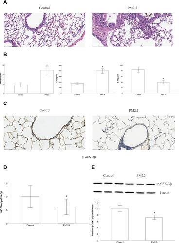
Figure 2 Effect of PM2.5 on HBEC viability.
Notes: CCK-8 assay results showed that cell viability did not markedly change after 24 h of PM2.5 (5–20 μg/mL) treatment, but cell viability decreased after treatment with 40 μg/mL PM2.5. #P<0.05, versus PM2.5 group, n=3.
Abbreviations: PM2.5, particulate matter<2.5 μm.
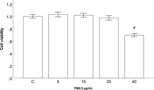
Figure 3 PM2.5 reduced the expression of p-GSK-3β in HBECs.
Notes: (A) Western blot analysis revealed that the expression of p-GSK-3β did not change markedly after 24 h of PM2.5 (5–10 μg/mL) treatment, whereas the expression was significantly decreased after PM2.5 stimulation at 20 μg/mL. (B) Western blot analysis showed that the expression of p-GSK-3β did not change markedly after 3 h of PM2.5 (20 μg/mL) treatment, but the expression was substantially decreased after 12 and 24 h of PM2.5 treatment. #P<0.05, compared to control group. n=3.
Abbreviations: PM2.5, particulate matter<2.5 μm.
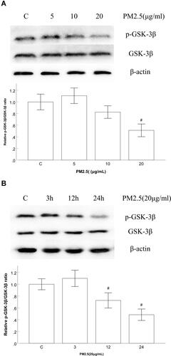
Figure 4 GSK-3β inhibitors increased the expression of p-GSK-3β in HBECs.
Notes: (A) Western blot analysis showed that the GSK-3β inhibitors TDZD-8 and SB216763 significantly increased the expression of p-GSK-3β. #P<0.05 compared to the control group. *P<0.05, compared to PM2.5 group, n=3.
Abbreviations: PM2.5, particulate matter<2.5 μm.
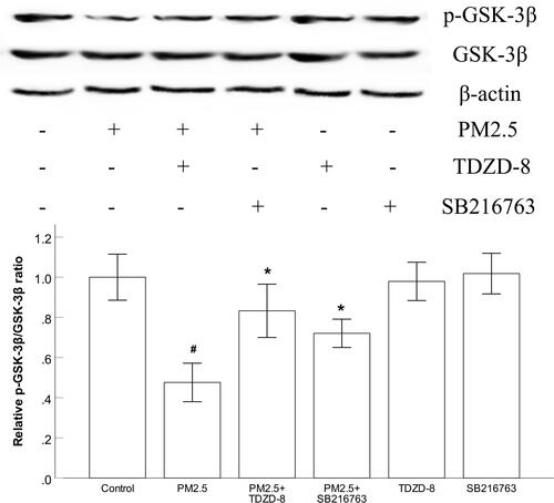
Figure 5 GSK-3β inhibitors alleviated PM2.5-activated JNK pathways in HBECs.
Notes: (A) Western blot analysis showed that the GSK-3β inhibitors TDZD-8 and SB216763 markedly decreased the PM2.5-induced ratio of p-JNK/JNK. #P<0.05, compared to control group. *P<0.05, compared to PM2.5 group, n=3.
Abbreviations: PM2.5, particulate matter<2.5 μm.
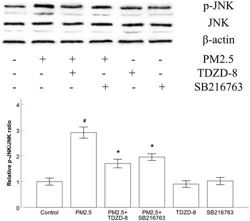
Figure 6 GSK-3β inhibitors and JNK inhibitors suppressed PM2.5-induced NF-κB activation and IkBα degradation in HBECs.
Notes: (A) Western blot analysis revealed that the GSK-3β inhibitors TDZD-8 and SB216763 markedly diminished PM2.5-induced NF-κB phosphorylation. (B) Western blot analysis showed that TDZD-8 and SB216763 markedly suppressed PM2.5-induced IκBα phosphorylation and IkBα degradation, respectively. (C) The JNK inhibitor SP600125 markedly abated the level of phosphorylated NF-κB induced by PM2.5. #P<0.05, compared to control group. *P<0.05, compared to PM2.5 group, n=3.
Abbreviations: PM2.5, particulate matter<2.5 μm.
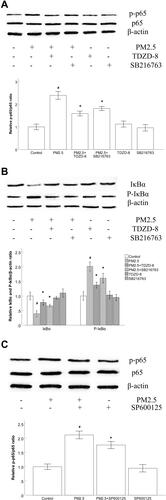
Figure 7 GSK-3β inhibitors blocked PM2.5-induced inflammatory responses in HBECs.
Notes: (A) The results of qRT–PCR showed that the GSK-3β inhibitors TDZD-8 and SB216763 markedly attenuated the production of HMGB1 and IL-6 induced by PM2.5. (B) ELISA results showed that the GSK-3β inhibitors TDZD-8 and SB216763 markedly prevented the production of HMGB1 induced by PM2.5. (C) ELISA results showed that TDZD-8 and SB216763 markedly prevented the production of IL-6 induced by PM2.5. #P<0.05, compared to control group. *P<0.05, compared to PM2.5 group, n=3.
Abbreviations: PM2.5, particulate matter<2.5 μm.
