Figures & data
Figure 1 Glipr1 knockout exacerbated CS-induced airway damage. (A) mRNA and (B) protein levels of Glipr1 in lung tissues of mice were increased after CS exposure. (C) Weight of mice (in grams) after CS exposure. (D) Hematoxylin and eosin (HE) staining. (E–H) Quantitative analysis of lung damage as assessed histopathologically. (E) Lung injury scores. (F) Mean linear intercept (MLI). (G) Mean alveolar septal thickness (MAST). (H) Destructive index (DI). Ten fields were randomly selected for scoring. (I) Masson’s staining. Error bars represent SD. *P < 0.05; **P < 0.01; ***P < 0.001.
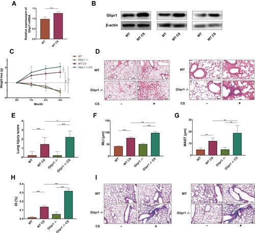
Figure 2 Glipr1 knockout caused more severe inflammatory infiltration after cigarette smoke exposure. (A) Total protein levels, (B) total cell counts, (C) neutrophils, (D) macrophages, and (E) lymphocytes in bronchoalveolar lavage fluid (BALF) were assessed. IL-6 concentrations in (F) BALF and (G) plasma samples were measured by ELISA. (H) MMP-9 concentration in BALF were measured by ELISA. (I) Ratio of MMP-9/TIMP-1 in the BALF. Error bars represent SD. *P < 0.05; **P < 0.01; ***P < 0.001.
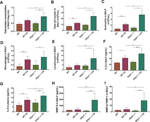
Figure 3 Glipr1 knockout aggravated caspase-1-dependent pyroptosis in CS-induced airway damage. (A) Levels of pro-caspase-1, cleaved-caspase-1, gasdermin D (GSDMD), GSDMD N-terminal domain, and β-actin proteins in the lung tissues of mice were measured by Western blotting analysis. Quantitative analysis of (B) cleaved-caspase-1 and (C) GSDMD N-terminal domain. (D) Levels of pro-IL-1β and cleaved- IL-1β in the lung tissues of mice were measured by Western blotting analysis. (E) Quantitative analysis of cleaved-IL-1β expression. (F) IL-1β concentration in bronchoalveolar lavage fluid (BALF) were measured by ELISA. Error bars represent SD. *P < 0.05; **P < 0.01; ***P < 0.001.
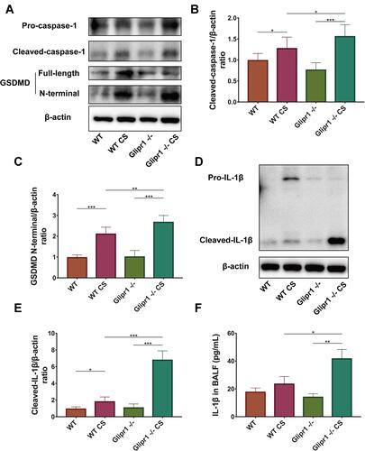
Figure 4 Glipr1 protected CS-induced airway damage through PLAU/EGFR signaling. (A) Levels of Glipr1, Plau, Egfr, p-Egfr, and β-actin proteins in the lung tissues of mice were measured by Western blotting analysis. Quantitative analysis of (B) Glipr1, (C) Plau, (D) Egfr, and (E) p-Egfr. (F) mRNA levels and (G) protein expression in bronchoalveolar lavage fluid (BALF) of Plau. (H) IHC. Error bars represent SD. *P < 0.05; **P < 0.01; ***P < 0.001.
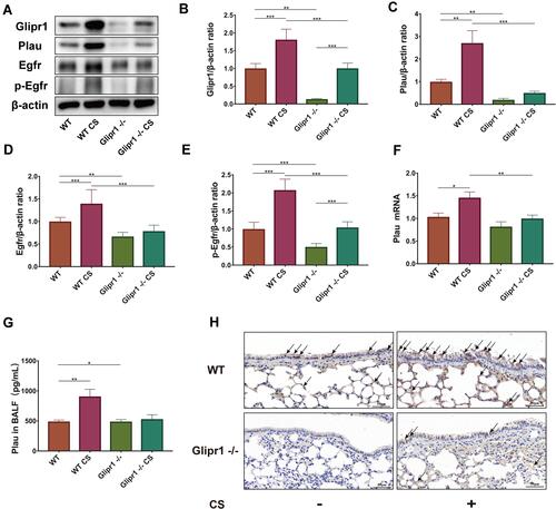
Figure 5 GLIPR1 knockdown exacerbated CSE-induced pyroptosis in vitro. (A) Levels of pro-caspase-1, cleaved-caspase-1, gasdermin D (GSDMD), GSDMD N-terminal domain, and β-actin proteins in human bronchial epithelial (HBE) cells were measured by Western blotting analysis. (B) Levels of pro-IL-1β and cleaved-IL-1β in HBE cells were measured by Western blotting analysis. shRNA vectors targeting GLIPR1.
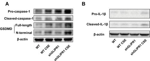
Figure 6 GLIPR1 knockdown reduced PLAU/EGFR signaling in vitro. (A) Levels of GLIPR1, PLAU, EGFR, p-EGFR, and β-actin proteins in human bronchial epithelial (HBE) cells were measured by Western blotting analysis. Quantitative analysis of (B) GLIPR1, (C) PLAU, (D) EGFR, and (E) p-EGFR. mRNA levels of (F) GLIPR1, (G) PLAU, (H) EGFR. (I) GLIPR1 was pull downed by Western blotting using anti- GLIPR1 antibody. (J) Immunoprecipitation assay showed that PLAU was pull downed by GLIPR1. (K) PLAU was pull downed by Western blotting using anti- PLAU antibody. (L) Immunoprecipitation assay showed that GLIPR1 was pull downed by PLAU. Error bars represent SD. *P < 0.05; **P < 0.01; ***P < 0.001. shRNA vectors targeting GLIPR1.
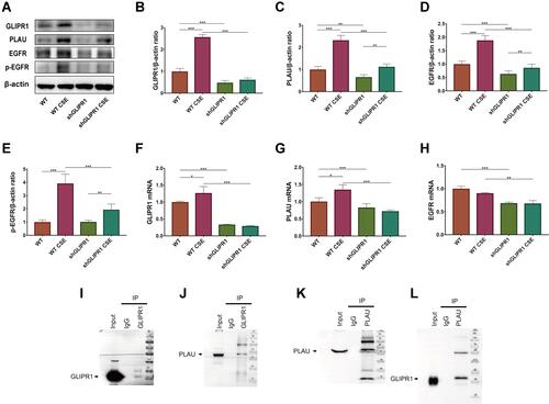
Figure 7 PLAU knockdown exacerbated CSE-induced pyroptosis in vitro. (A) Levels of pro-caspase-1, cleaved-caspase-1, gasdermin D (GSDMD), GSDMD N-terminal domain, and β-actin proteins in human bronchial epithelial (HBE) cells were measured by Western blotting analysis. (B) Levels of pro-IL-1β and cleaved-IL-1β in HBE cells were measured by Western blotting analysis. shRNA vectors targeting PLAU.
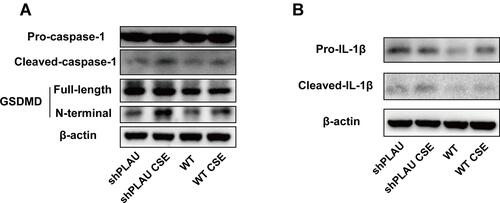
Figure 8 PLAU knockdown reduced EGFR signaling in vitro. (A) Levels of PLAU, EGFR, p-EGFR, and β-actin proteins in human bronchial epithelial (HBE) cells were measured by Western blotting analysis. Quantitative analysis of (B) PLAU, (C) EGFR, and (D) p-EGFR. mRNA levels of (E) PLAU, (F) EGFR. Error bars represent SD. *P < 0.05; **P < 0.01; ***P < 0.001. shRNA vectors targeting PLAU.
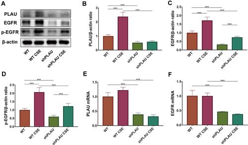
Figure 9 PLAU overexpression reversed CSE-induced pyroptosis in vitro. (A) Levels of pro-caspase-1, cleaved-caspase-1, gasdermin D (GSDMD), GSDMD N-terminal domain, and β-actin proteins in human bronchial epithelial (HBE) cells were measured by Western blotting analysis. (B) Levels of pro-IL-1β and cleaved-IL-1β in HBE cells were measured by Western blotting analysis. Quantitative analysis of (C) cleaved-caspase-1, (D) GSDMD N-terminal domain, and (E) cleaved- IL-1β. Error bars represent SD. *P < 0.05; **P < 0.01; ***P < 0.001.
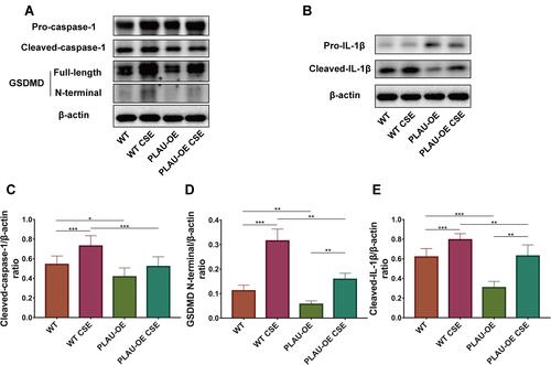
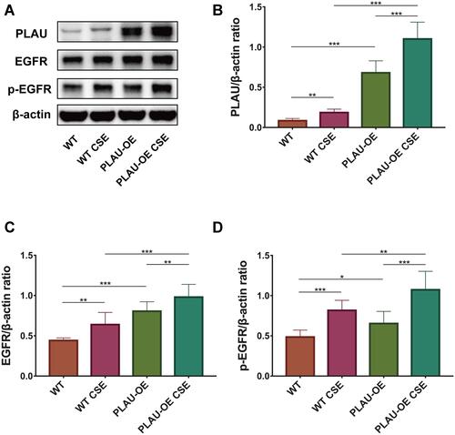
Figure 10 PLAU overexpression increased EGFR signaling in vitro. (A) Levels of PLAU, EGFR, p-EGFR, and β-actin proteins in human bronchial epithelial (HBE) cells were measured by Western blotting analysis. Quantitative analysis of (B) PLAU, (C) EGFR, and (D) p-EGFR. Error bars represent SD. *P < 0.05; **P < 0.01; ***P < 0.001.