Figures & data
Figure 3 Detection of airway fibrosis.
Notes: (A) Con group. (B) COPD group. (C) COPD+AE group. (D) Detection of collagen fiber deposition in the airway wall. The airway collagen increased after LPS administration, while exercise administration decreased fibrotic score. The results were expressed as μm2 of collagen fibers per μm2 of tissue area. #P<0.05 compared with the Con group. *P<0.05 compared with the COPD group (magnification, x40).
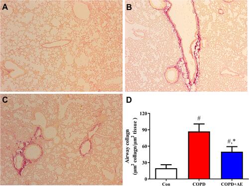
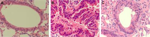
Figure 7 Detection of bronchial mucus cell hyperplasia and bronchoconstriction.
Notes: (A) Con group. (B) COPD group. (C) COPD+AE group. LPS administration led to severe bronchial mucus cell hyperplasia and bronchoconstriction, while 4 weeks of AE improved bronchial mucus cell hyperplasia and bronchoconstriction (magnification, x400).