Figures & data
Table 1 Clinical Evaluation Outcomes at Baseline and 3 Months After Endobronchial Occlusion Using the Silicone Plugs
Figure 1 Procedure of the bronchoscopic silicone plug placement. (A) The customized silicone plug which we used in the study. (B) Preoperative planning was designed via the virtual bronchoscopic navigation system. (C) The tip knot of the plug was grasped by the grasping forceps to guide the route and adjust orientation. (D) The silicone plug was placed into the targeted bronchi by flexible bronchoscopy.
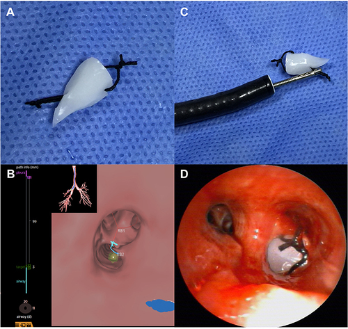
Figure 2 Individual results at baseline and 3 months after the bronchoscopic silicone plug placement.
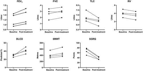
Figure 3 Chest CT scan images at baseline, 7 days and 3 months after the bronchoscopic silicone plug placement in Case 1. (A) A GEB occupying almost entirely the right upper lobe before treatment. (B) At 7 days after the procedure, a CT scan showed complete silicone plugs placement of the GEB and re-expansion of the adjacent lung. (C) The radiological improvement was sustained for 3 months after treatment.
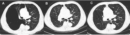
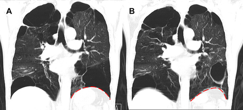
Figure 4 Coronal CT scan images at baseline and 3 months after the bronchoscopic silicone plug placement in Case 2. (A) Preoperative chest CT scan showed a GEB located in the left lower lobe. (B) Three months after the procedure, repeated CT demonstrated regression of the GEB with the upshift of the diaphragm without the collapse of the GEB. The red dotted line represents the shape of diaphragm.