Figures & data
Table 1 Characteristics of patients with and without pulmonary congestion at baseline, assessed by standardized and routine procedure
Figure 1 Randomly selected chest radiographs assessed negative in the routine assessment and positive in the standardized assessment.
Abbreviations: FEV1, forced expiratory volume in 1 second; FVC, forced vital capacity; NT-proBNP, N-terminal prohormone of brain natriuretic peptide.
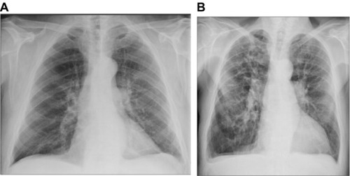
Figure 2 ROC curves for the routine (red dashed) and standardized (blue) assessments.
Notes: Age-adjusted logrank P<0.0001 between R−/S− and R−/S+; age-adjusted P=0.049 between R+/S− and R−/S+. The results were based on the 99 index admissions.
Abbreviations: COPD, chronic obstructive pulmonary disease; R−, radiological assessment negative; R+, radiological assessment positive; S−, standardized assessment negative; S+, standardized assessment positive; ROC, receiver-operating characteristics.
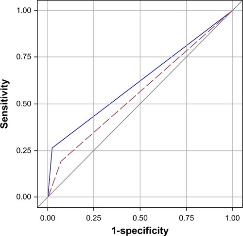
Figure 3 Survival after acute exacerbation of COPD, stratified by radiologic evaluation of pulmonary congestion on admission.
Abbreviations: AUC, area under the curve; ROC, receiver operating characteristics.
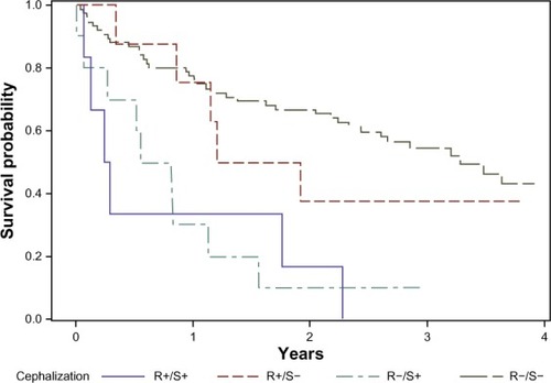
Table 2 Number of mortalities (with mortality rates per 100 patient-years) in patients with AECOPD, by selected dichotomous covariables
Table 3 Number of mortalities (with mortality rates per 100 patient-years) in patients with AECOPD, by quartiles of selected continuous covariables
Table 4 Hazard ratios (with 95% confidence intervals) for dying during a median of 1.9 years follow up after acute exacerbation of COPD
Table 5 Geometric mean (with number of observations) of baseline NT-proBNP concentration (pg/mL), by selected dichotomous variables
Table 6 Geometric mean of baseline NT-proBNP concentrations (pg/mL), by quartiles of selected continuous variables
Table 7 Relative concentration of NT-proBNP in patients with acute exacerbation of COPD