
Figures & data
Figure 1 Radiographic image of the area displaced by diaphragm motion during inspiration and expiration for the same chronic obstructive pulmonary disease patient.
Note: The thick horizontal lines of posteroanterior radiographs represent the costal insertion of diaphragm to the same spinal process of vertebral column between full inspiration and full expiration.
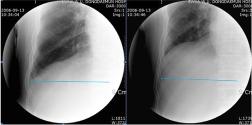
Figure 2 Schematic illustration of the area displaced by diaphragm movement during both inspiration (Area I) and expiration (Area E) in patients with chronic obstructive pulmonary disease.
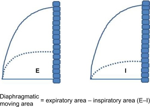
Table 1 Subject characteristics of chronic obstructive pulmonary disease patients (N=37)
Table 2 Anthropometric and pulmonary function data (n=37)
Table 3 Results of pulmonary function test between pre- and postpulmonary rehabilitation in patients with chronic obstructive pulmonary disease (n=37)
Table 4 Changes of diaphragmatic area between expiratory–inspiratory diaphragm motion with fluoroscopy in patients with chronic obstructive pulmonary disease (n=37)