Figures & data
Figure 1 Chest computed tomography scan. Coronal (left) and horizontal (right) images show severe bronchiectasis with large cysts and mucus plugging.
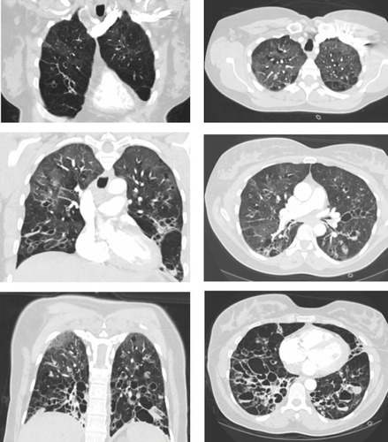
Figure 2 Capillary zone electrophoresis. A decrease of the alpha-1 globin peak is notable.
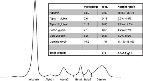
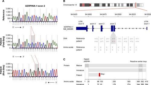
Figure 3 Sequence of a regular and mutated SERPINA-1 gene. (A) DNA sequencing chromatogram of reference sequence (upper) and patient sequence (middle and lower). (B) Overview of SERPINA-1 gene location, structure, and patient’s mutation with DNA and amino acid sequence. (C) Overview of mature, immature, and patient SERPINA-1 protein and domains.
Abbreviations: Fwd-Seq, forward sequence; Rvs-Seq, reverse sequence; UTR, untranslated region.