
Figures & data
Figure 1 Application of the proposed algorithms on the CT scans of a COPD patient.
Abbreviations: COPD, chronic obstructive pulmonary disease; CT, computed tomography.
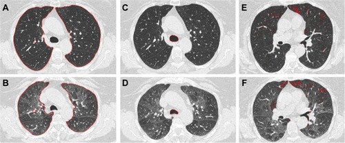
Table 1 Anthropometric data, smoke exposure, pulmonary function, and presence of purulent sputum in 69 patients with COPD subdivided according to the predominant mechanism of air flow limitation and sex
Table 2 CT quantitative densitometric data, airway wall thickness, inspiratory–expiratory volume variation, and tracheal collapsibility in 69 patients with COPD subdivided according to the predominant mechanism of air flow limitation and sex
Table 3 Relationships of intrathoracic tracheal collapsibility with functional parameters indicative of airflow obstruction and hyperinflation
Table 4 Relationships of intrathoracic tracheal collapsibility with CT thresholds of lung inspiratory and expiratory density (%LAA−950insp and %LAA−910exp, respectively)
Figure 2 Relationship of intrathoracic tracheal collapsibility with delta volume ([inspiratory CT volume – expiratory CT volume]/inspiratory CT volume) in 69 patients with COPD (A); 28 of whom were classified as being affected by predominant airway disease (B); 41 of whom were classified as being affected by predominant emphysema (C); males (D); and females (E).
![Figure 2 Relationship of intrathoracic tracheal collapsibility with delta volume ([inspiratory CT volume – expiratory CT volume]/inspiratory CT volume) in 69 patients with COPD (A); 28 of whom were classified as being affected by predominant airway disease (B); 41 of whom were classified as being affected by predominant emphysema (C); males (D); and females (E).](/cms/asset/2f0bc97d-2f8f-4747-b28f-48e5457dfe02/dcop_a_80558_f0002_b.jpg)
Figure 3 Intrathoracic tracheal collapsibility in males and females with COPD according to predominant airway disease and predominant emphysema.
Abbreviation: COPD, chronic obstructive pulmonary disease.
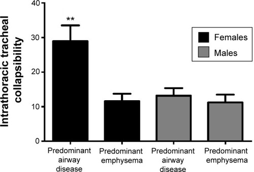
Table 5 Predictive models of intrathoracic tracheal collapsibility by multivariate regression analysis of clinical history data (cough, wheezing, dyspnea), pulmonary function, and CT variables in the whole set of patients with COPD and in the examined subsets