Figures & data
Table 1 Demographic detail and lung function data of CAL patients and control subjects
Table 2 Comparison of different pathological indices in CAL large and small airways
Table 3 Comparison of different pathological indices in CAL small airway and normal control small airway
Figure 1 Large airway tissue from patient with CAL.
Notes: Black arrows indicate large-airway epithelium (chronic airflow limitation) stained positive for EGFR (epithelial activation marker). Original magnification, ×360. Scale bar =50 µm.
Abbreviations: CAL, chronic airflow limitation; EGFR, epidermal growth factor receptor.
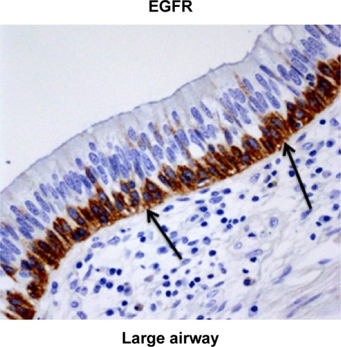
Figure 2 (A) Percentage of large-airway epithelium in CAL patients stained for EGFR compared with CAL small airway and normal control small airway. (B) Percentage of Rbm fragmentation (total length of clefts as percentage of the total length of Rbm) in large airway in CAL patients versus CAL small airway and in normal control small airway.
Abbreviations: CAL, chronic airflow limitation; EGFR, epidermal growth factor receptor; Rbm, reticular basement membrane.
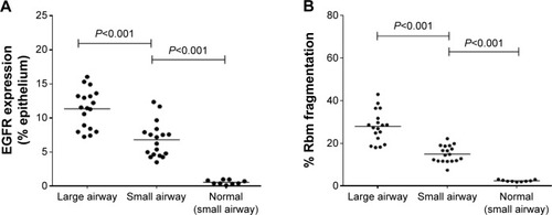
Figure 3 Comparison of small airway in CAL subjects with normal control.
Abbreviations: CAL, chronic airflow limitation; EGFR, epidermal growth factor receptor; Rbm, reticular basement membrane.
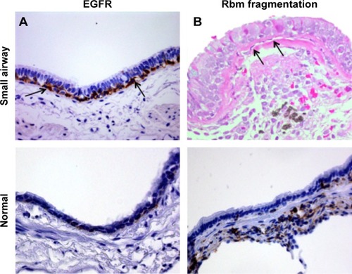
Figure 4 Large-airway tissue from patient with CAL.
Abbreviations: CAL, chronic airflow limitation; EMT, epithelial mesenchymal transition; Rbm, reticular basement membrane.
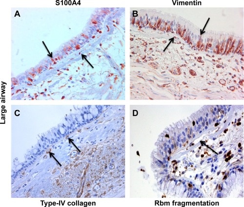
Figure 5 (A) Comparison of S100A4 expression in the (basal) epithelial cells of large airway in CAL patients, with small airway of CAL patients and normal control small airway. (B) Comparison of S100A4 expression in Rbm of large airway in CAL patients with small airway of CAL patients and normal control small airway.
Abbreviations: CAL, chronic airflow limitation; Rbm, reticular basement membrane.
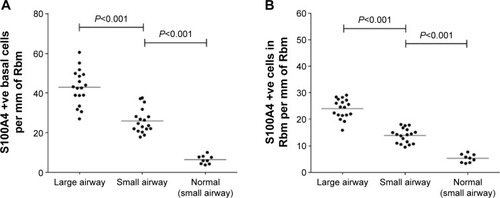
Figure 6 (A) Comparison of number of basal epithelial cells positive for vimentin in CAL large airway versus CAL small airways and in normal control small airway. (B) Comparison of number of vimentin-positive cells in Rbm in CAL large airway versus CAL small airway and normal control small airway.
Abbreviations: CAL, chronic airflow limitation; Rbm, reticular basement membrane.
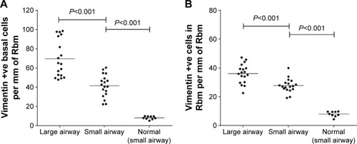
Figure 7 Small airway specimens from patients with CAL stained for EMT biomarkers and vascularity marker, compared with normal control small airway for comparison.
Abbreviations: CAL, chronic airflow limitation; EMT, epithelial mesenchymal transition; Rbm, reticular basement membrane.
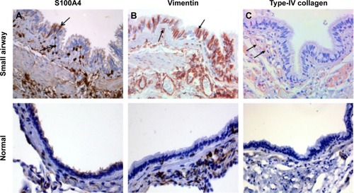
Figure 8 Number of vessels in Rbm in large airway of CAL compared with CAL small airway and normal control small airway.
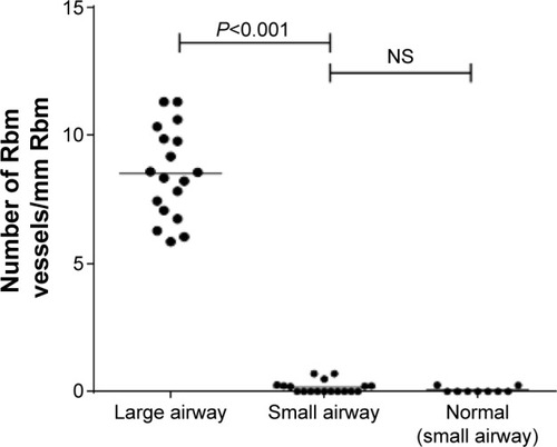
Table 4 Linear regression of pathological data for large and small airways from CAL subjects versus spirometric indices of airflow obstruction
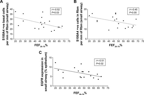
Figure 9 (A) Correlation between the number of small airway basal epithelial cells (per millimeter of Rbm) that were positive for S100A4 with FEF25–75% (an index of small airway caliber). (B) Correlation between the number of S100A4-positive cells in Rbm (per millimeter of Rbm) in small airway and FEF25–75%. (C) Correlation between EGFR (% epithelium) in small airway and FEF25–75%.