Figures & data
Figure 1 HRCT images of the case.
Abbreviation: HRCT, high-resolution computed tomography.
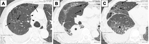
Figure 2 Gross appearance of the right lung.
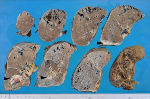
Figure 3 Computed tomography images and their corresponding histological images of right lower lobe.
Notes: (A) Computed tomography image shows coarse reticulation with honeycomb-like clustered cystic airspaces. Squared area corresponds to microscopic observation (C). (B) A gross picture of the same lesion. Squared area corresponds to microscopic observation (C). (C) Microscopic observation revealed cystic spaces and interstitial fibrosis. Arrowheads indicate areas corresponding to honeycomb cysts.
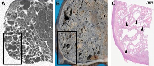
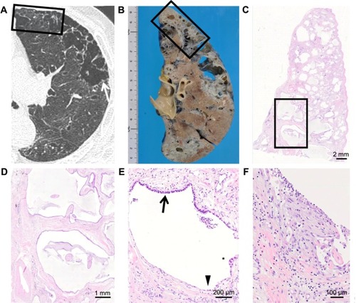
Figure 4 Computed tomography images and their corresponding histological images of right upper lobe.
Notes: (A) Computed tomography image shows subtle fine reticulation (arrow) which can be identified with careful observations. (B) A gross picture of the same area. (C) Microscopic observation of squared area in (A, B). (D) Close-up of the squared area in (C) revealed significant interstitial fibrosis.
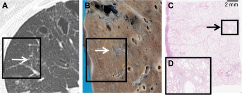
Figure 5 Computed tomography images and their corresponding histological images of left upper lobe.