
Figures & data
Table 1 Brain regions exhibiting 24-hour rhythms in vivo
Figure 1 Phase shift in the diurnal rhythm of SPK.
Notes: (A and B) Chronic tracking of SPK rates for preinjury, latency, and spontaneously seizing stages of epileptogenesis for a single rat. Dot-dashed vertical lines correspond to midnight. (C) Time series were combined into a 24-hour time windows (modulo 24) revealing a post-injury phase shift that occurs during the latency period, before spontaneous seizures appear. (D) Circadian rhythm of CBT are shown. There was no observed phase shift after injury, although the variability of the signal did increase. Copyright © 2013, The American Physiological Society. Adapted from Stanley DA, Talathi SS, Parekh MB, et al. Phase shift in the 24-hour rhythm of hippocampal EEG spiking activity in a rat model of temporal lobe epilepsy. J Neurophysiol. 2013;110(5):1070–1086.Citation66
Abbreviations: CBT, core body temperature; h, hours; SPK, hippocampal spiking activity; SS, spontaneously seizing.
Abbreviations: CBT, core body temperature; h, hours; SPK, hippocampal spiking activity; SS, spontaneously seizing.
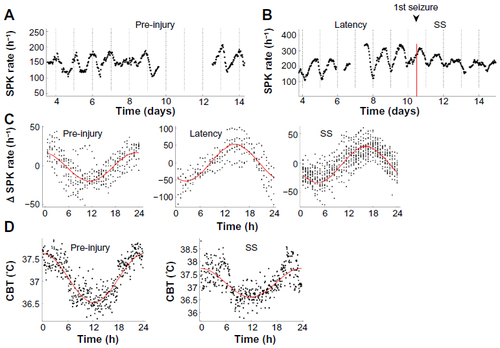
Table 2 Half-lives of common antiepileptic drugs