Figures & data
Figure 1 Spectrum of extracellular matrices.
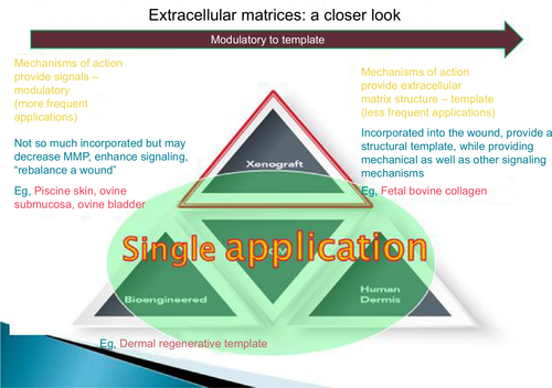
Figure 2 Increased acute wound closure rate seen with AFS over SIS in an acute wound care model.
Abbreviations: AFS, acellular fish skin; SIS, small intestine submucosa.
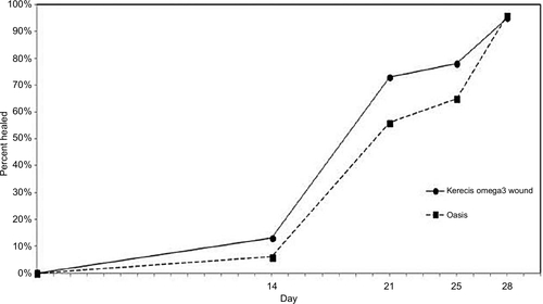
Figure 3 Increased acute wound closure rate seen with AFS over dHACM in an acute wound care model.
Abbreviations: AFS, acellular fish skin; dHACM, dehydrated human amnion/chorion membrane.
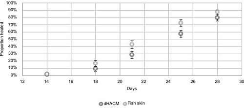
Figure 4 Efficacy of AFS as a bacterial barrier and the increased efficacy seen with the addition of greater omega-3 fatty acids.
Abbreviation: AFS, acellular fish skin.
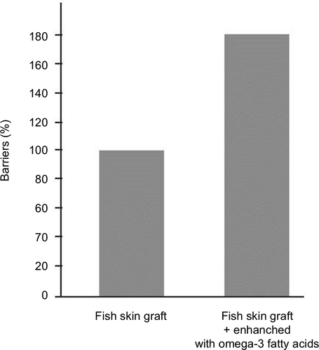
Figure 5 Clinical use of antibiotics before and after the application of AFS in a cohort of chronic wound patients.
Abbreviation: AFS, acellular fish skin.
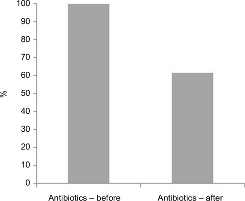
Table 1 Studies evaluating the use of AFS in wound healing
Figure 6 Mean wound surface area as noted during the 5-week AFS treatment and 3-week follow-up of venous leg ulcers.Citation11
Abbreviation: AFS, acellular fish skin.
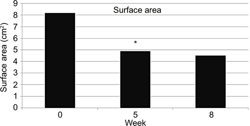
Table 2 Likelihood of healing by prognostic factors
Figure 7 (A) Post-debrided foot with exposed bone. (B) Completion of transmetatarsal amputation. (C) Placement of AFS. (D) Post-op day 4 with no negative-pressure wound therapy. (E) Three weeks after grafting.
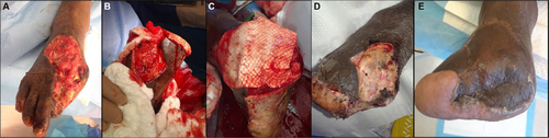
Figure 8 Levels of omega-3 and omega-6 fatty acids in various products with y-axis being the percent of fatty acid and x-axis being the type of fatty acid.
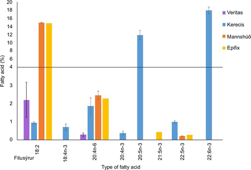
Table 3 Porcine burn model treatment groups
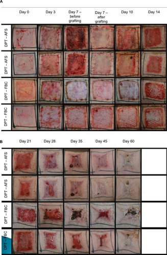
Figure 9 Progression of porcine wound closure when treated with AFS with and without skin grafting.