Figures & data
Figure 1 A chronic head wound with significant exudate in a neonate with JEB-GS.
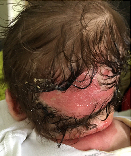
Figure 2 Right hand of a neonate with JEB-GS with thickened, dystrophic nails with periungual swelling present since birth.
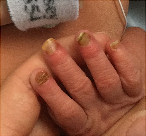
Figure 3 Right hand with new and some healing haemorrhagic blisters in a 6-year-old child with JEB-GI.
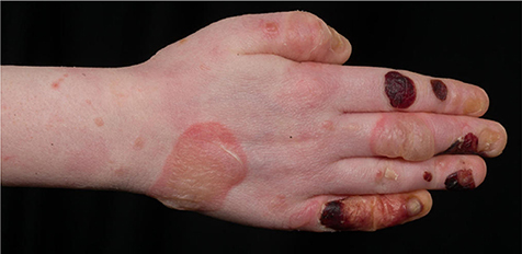
Figure 4 Chronic nail bed inflammation in a 6-year-old child with LOC Syndrome.
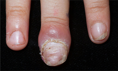
Figure 5 Chronic nappy wound on an infant with JEB-GS.
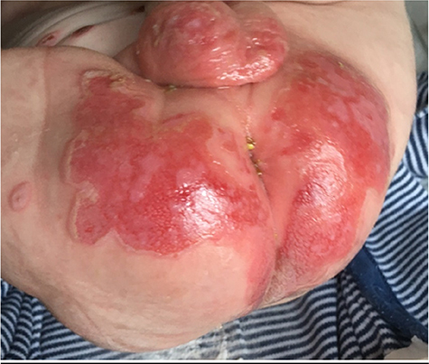
Figure 6 Chronic facial wound in a child with LOC syndrome.
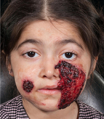
Figure 7 Tracheostomy with a chronic wound around the stoma site in a patient with LOC syndrome.
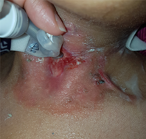
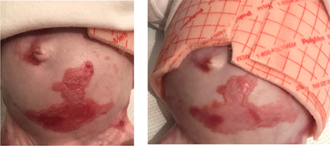
Figure 8 Chronic abdominal wound on infant with JEB-GS, demonstrating pre topical gentamicin use (left figure) with improvements after use (right figure).