Figures & data
Table 1 Mean extension angles between tibia and femur for both legs of CCH-injected and buffer-injected rats, with muscle on and muscle off and at four torques
Table 2 Statistical linear mixed effect modeling to determine the effect of CCH injection compared to buffer injection on the ROM of a rat knee with contracture
Figure 1 Pictures of the automatic arthrometer developed by The Bone and Joint Laboratory and used to measure the knee ROM in extension.
Abbreviation:DDDT_A_12181558_F0001
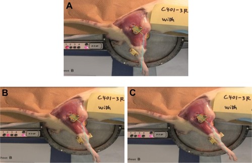
Figure 2 Mean extension angles of CCH-injected and buffer-injected experimental rat knees.
Abbreviations: CCH, collagenase Clostridium histolyticum; ROM, range of motion.
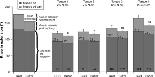
Figure 3 Box plots showing the degree of variation for ROM for experimental and contralateral knees of buffer-injected and CCH-injected rats.
Abbreviations: CCH, collagenase Clostridium histolyticum; ROM, range of motion.
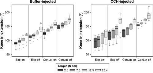
Figure 4 Immunostaining of collagen type I and collagen type III in the posterior capsule from contralateral knees and experimental knees intra-articularly injected with CCH or buffer.
Abbreviation: ns, not significant.
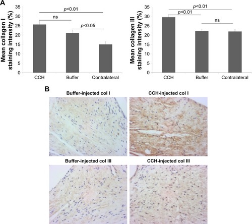
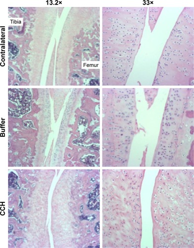
Figure 5 Hematoxylin and eosin staining of contralateral knees and experimental knees intra-articularly injected with CCH or buffer.
Abbreviation: CCH, collagenase Clostridium histolyticum.