Figures & data
Figure 1 Effects of agomelatine on oxidative stress induced by Aβ25–35 in PC12 cells.
Notes: (A, B) Measurement of cell viability, MDA, and LDH. PC12 cells were preincubated with or without agomelatine for 24 hours and then incubated with Aβ25–35 (20 µM) for 24 hours. (C) Measurement of ROS via flow cytometry analysis. PC12 cells were preincubated with or without agomelatine (2, 20, and 200 µM) for 24 hours and then incubated with Aβ25–35 (20 µM) for 24 hours. (D) Measurement of ROS via flow cytometry analysis. After Aβ25–35 pretreatment for 24 hours, PC12 cells were then exposed to agomelatine for 24 hours. Data represent the mean ± SEM of three independent experiments (n=3). *P<0.05 vs control; **P<0.01 vs control; #P<0.05 vs Aβ25–35 group; ##P<0.01 vs Aβ25–35 group.
Abbreviations: Aβ25–35, amyloid beta 25–35; Ago, agomelatine; CCK-8, cell counting kit-8; DCFH-DA, dichlorodihydrofluorescein diacetate; LDH, lactate dehydrogenase; MDA, malondialdehyde.
Abbreviations: Aβ25–35, amyloid beta 25–35; Ago, agomelatine; CCK-8, cell counting kit-8; DCFH-DA, dichlorodihydrofluorescein diacetate; LDH, lactate dehydrogenase; MDA, malondialdehyde.
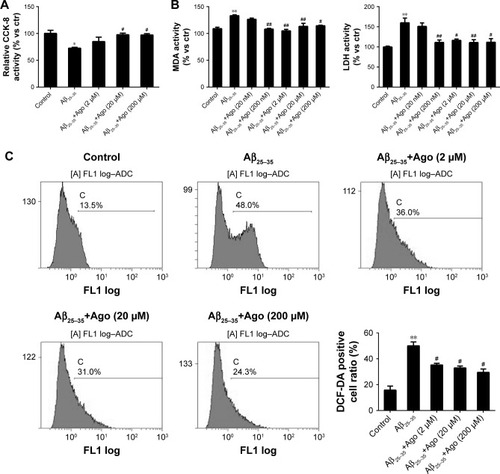
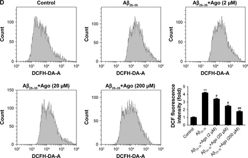
Figure 2 Effects of agomelatine on tau phosphorylation induced by Aβ25–35 in PC12 cells.
Notes: Measurement of P-tau and tau expression by Western blot in PC12 cells. PC12 cells were preincubated with or without agomelatine (20 µM) for 24 hours and then incubated with Aβ25–35 (20 µM) for 24 hours. Data represent the mean ± SEM of three independent experiments (n=3). *P<0.05 vs control; #P<0.05 vs Aβ25–35 group.
Abbreviations: Aβ25–35, amyloid beta 25–35; Ago, agomelatine.
Abbreviations: Aβ25–35, amyloid beta 25–35; Ago, agomelatine.
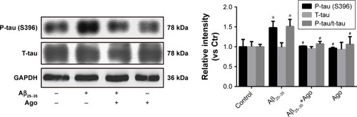
Figure 3 Effect of agomelatine and luzindole on oxidative stress and tau phosphorylation induced by Aβ25–35 in PC12 cells.
Notes: (A, C) Measurement of MDA, LDH, and ROS. PC12 cells untreated or treated with agomelatine (20 µM) for 24 hours in the absence or presence of luzindole (1 µM) for 24 hours were exposed to Aβ25–35 (20 µM) for 24 hours. (B) Measurement P-tau and tau expression by Western blot. PC12 cells untreated or treated with agomelatine (20 µM) for 24 hours in the absence or presence of luzindole (1 µM) for 24 hours were exposed to Aβ25–35 (20 µM) for 24 hours. Data represent the mean ± SEM of three independent experiments (n=3). *P<0.05; **P<0.01.
Abbreviations: Aβ25–35, amyloid beta 25–35; Ago, agomelatine; DCFH-DA, dichlorodihydrofluorescein diacetate; LDH, lactate dehydrogenase; MDA, malondialdehyde.
Abbreviations: Aβ25–35, amyloid beta 25–35; Ago, agomelatine; DCFH-DA, dichlorodihydrofluorescein diacetate; LDH, lactate dehydrogenase; MDA, malondialdehyde.
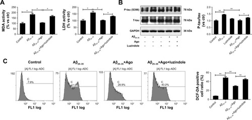
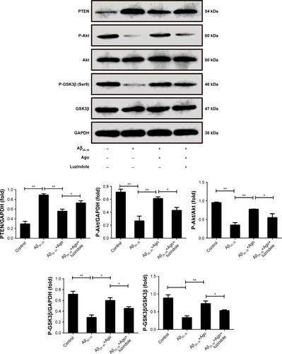
Figure 4 The signaling involved in the neuroprotective effect of agomelatine. Measurement of PTEN, P-Akt, Akt, P-GSK3β (Ser9), and GSK3β expression by Western blot in PC12 cells.
Notes: PC12 cells untreated or treated with agomelatine (20 µM) for 24 hours in the absence or presence of luzindole (1 µM) for 24 hours were exposed to Aβ25–35 (20 µM) for 24 hours. Data represent the mean ± SEM of three independent experiments (n=3). *P<0.05, **P<0.01.
Abbreviations: Aβ25–35, amyloid beta 25–35; Ago, agomelatine.
Abbreviations: Aβ25–35, amyloid beta 25–35; Ago, agomelatine.