Figures & data
Table 1 WHO listing (third revision, 2012)Citation16 of critically important antimicrobials for human medicine
Figure 1 Checkerboard magnetic chip for cell capture.
Notes: (A) Checkerboard magnetic array with alternating polarity. (B) Magnetic field strength (Bz) between two checkerboard arrays. (C) Magnetic force simulation for 1 µm magnetic beads exposed to a simple two-pole system or checkerboard array. (D) Chip-array arrangement. (E) The effect of flow rate on the enrichment ratio. (F) Magnetic enrichment of hybridoma cells. Cells were labeled with CD45-specific magnetic beads; IgG2b beads were used as a control. Image reproduced with permission from Park KS, Kim H, Kim S, et al. Nanomagnetic system for rapid diagnosis of acute infection. ACS Nano. 2017;11(11):11425–11432. Copyright 2017 American Chemical Society.Citation47
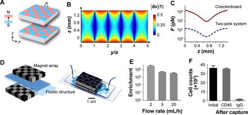
Figure 2 NP-coating attenuates Helicobacter pylori (H. pylori) pathobiology. Gastric cancer cells were infected with NP -H. pyloriGFP complexes estimated to maximally cover approximately 25% (Si30–25%: 1 × 10⁸ bacteria, 600 µg mL−1 Si30; 10 min PBS) or 0.25% (Si30–0.25%: 1 × 10⁸ bacteria, 60 µg mL−1 Si30, 10 min PBS) of the bacterial surface, respectively.
Notes: (A) Fluorescent microscope image demonstrating NP inhibition of the “hummingbird” phenotype. (B) Immunoblot analysis showing NP (Si30) attenuated type IV secretion system function and decreasing phosphorylated CagA levels. (C) β-Actin normalized phosphorylated CagA levels. (D) NP concentration-dependent decrease in IL-8 secretion. Westmeier D, Posselt G, Hahlbrock A, et al. Nanoparticle binding attenuates the pathobiology of gastric cancer-associated Helicobacter pylori. Nanoscale. 2018;10(3):1453–1463. Adapted by permission of The Royal Society of Chemistry.Citation95 Statistical significance was determined by using the Mann-Whitney test or paired t-test assuming significance at *P=0.05.
Abbreviations: Ctrl, control; NP, nanoparticle.
Abbreviations: Ctrl, control; NP, nanoparticle.
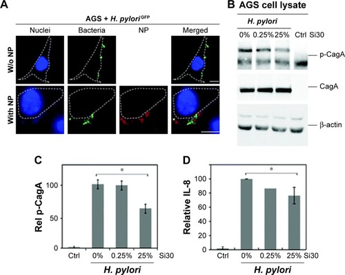
Figure 3 In vivo anti-Helicobacter pylori therapeutic efficacy of AGS-NP (CLR).
Notes: (A) H. pylori infection mouse model subject to inoculation, development, and treatment protocols. (B) Analysis of bacterial loads in mice treated with PBS, free CLR, PEG-NP (CLR), or AGS-NP (CLR) (n=6 per group). Bars represent median values. *P<0.05, **P<0.01, and ***P<0.001. Image reproduced from Coating nanopar-ticles with gastric epithelial cell membrane for targeted antibiotic delivery against Helicobacter pylori infection. Advanced Therapeutics. 2018;1(2):1800016. Copyright Wiley-VCH Verlag GmbH & Co. KGaA. Reproduced with permission.Citation126
Abbreviations: CFU, colony-forming unit; CLR, clarithromycin; NP, nanoparticle.
Abbreviations: CFU, colony-forming unit; CLR, clarithromycin; NP, nanoparticle.
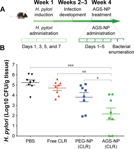
Table 2 Targeted antibacterial therapies
Figure 4 Structure of hNS.
Notes: (A) Schematic diagram depicting hNS and its mechanism of neutralizing PFTs. (B) Nanoparticle hydrodynamic size (diameter) and zeta potential (mV) (n=3) before and after membrane coating. (C) Transmission electron microscopy image of hNS. (D) Diameter stability of hNS over 5 days in different media (n=3). Image modified with permission from Chen Y, Chen M, Zhang Y, et al. Broad-spectrum neutralization of pore-forming toxins with human erythrocyte membrane-coated nanosponges. Adv Healthc Mater. 2018;7(13):e1701366. © 2018 WILEY-VCH Verlag GmbH & Co. KGaA, Weinheim.Citation155
Abbreviations: hNS, human RBC nanosponge; PFTs, pore-forming toxins; PLGA, poly (lactide-co-glycolide); RBC, red blood cell.
Abbreviations: hNS, human RBC nanosponge; PFTs, pore-forming toxins; PLGA, poly (lactide-co-glycolide); RBC, red blood cell.
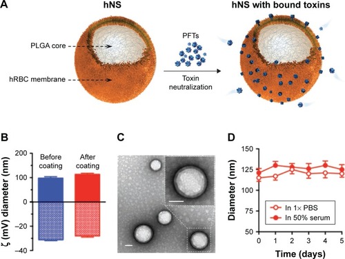
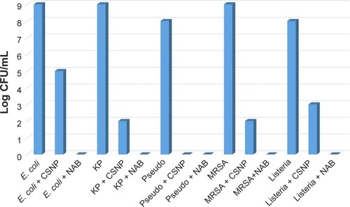
Figure 5 CFU assay.
Note: Image reproduced from Jamil et alCitation181 with permission (Copyright 2016 Frontier Publishing).
Abbreviations: CFU, colony-forming unit; CSNP, chitosan nanoparticles; E. coli, Escherichia coli; KP, Klebsiella pneumoniae; MRSA, methicillin-resistant Staphylococcus aureus; NAB, nano-antibiotic; Pseudo, Pseudomonas aeruginosa.
Abbreviations: CFU, colony-forming unit; CSNP, chitosan nanoparticles; E. coli, Escherichia coli; KP, Klebsiella pneumoniae; MRSA, methicillin-resistant Staphylococcus aureus; NAB, nano-antibiotic; Pseudo, Pseudomonas aeruginosa.